 Movie
Movie Controller
 Structure viewers
 About Yorodumi Papers


+Search query
-Structure paper
| Title | Solution structure of the 128 kDa enzyme I dimer from Escherichia coli and its 146 kDa complex with HPr using residual dipolar couplings and small- and wide-angle X-ray scattering. |
|---|---|
| Journal, issue, pages | J Am Chem Soc, Vol. 132, Issue 37, Page 13026-13045, Year 2010 |
| Publish date | Sep 22, 2010 |
 Authors Authors | Charles D Schwieters / Jeong-Yong Suh / Alexander Grishaev / Rodolfo Ghirlando / Yuki Takayama / G Marius Clore /  |
| PubMed Abstract | The solution structures of free Enzyme I (EI, ∼128 kDa, 575 × 2 residues), the first enzyme in the bacterial phosphotransferase system, and its complex with HPr (∼146 kDa) have been solved using ...The solution structures of free Enzyme I (EI, ∼128 kDa, 575 × 2 residues), the first enzyme in the bacterial phosphotransferase system, and its complex with HPr (∼146 kDa) have been solved using novel methodology that makes use of prior structural knowledge (namely, the structures of the dimeric EIC domain and the isolated EIN domain both free and complexed to HPr), combined with residual dipolar coupling (RDC), small- (SAXS) and wide- (WAXS) angle X-ray scattering and small-angle neutron scattering (SANS) data. The calculational strategy employs conjoined rigid body/torsion/Cartesian simulated annealing, and incorporates improvements in calculating and refining against SAXS/WAXS data that take into account complex molecular shapes in the description of the solvent layer resulting in a better representation of the SAXS/WAXS data. The RDC data orient the symmetrically related EIN domains relative to the C(2) symmetry axis of the EIC dimer, while translational, shape, and size information is provided by SAXS/WAXS. The resulting structures are independently validated by SANS. Comparison of the structures of the free EI and the EI-HPr complex with that of the crystal structure of a trapped phosphorylated EI intermediate reveals large (∼70-90°) hinge body rotations of the two subdomains comprising the EIN domain, as well as of the EIN domain relative to the dimeric EIC domain. These large-scale interdomain motions shed light on the structural transitions that accompany the catalytic cycle of EI. |
 External links External links | J Am Chem Soc / PubMed:20731394 / PubMed Central |
| Methods | SAS (X-ray synchrotron) / NMR (solution) / small angle scattering |
| Structure data | 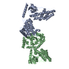 SASDCN3:  PDB-2kx9: 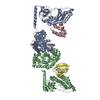 PDB-2xdf: |
| Source |
|
 Keywords Keywords |  TRANSFERASE / SUGAR PHOSPHOTRANSFERASE SYSTEM / PTS / SUGAR TRANSPORT TRANSFERASE / SUGAR PHOSPHOTRANSFERASE SYSTEM / PTS / SUGAR TRANSPORT |

