 Movie
Movie Controller

[English] 日本語
 Yorodumi
- PDB-2kx9: Solution Structure of the Enzyme I dimer Using Residual Dipolar C... -
+ Open data

- Basic information
| Entry | Database: PDB / ID: 2kx9 | ||||||
|---|---|---|---|---|---|---|---|
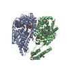
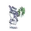
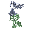
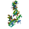
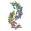
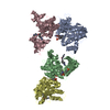
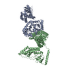
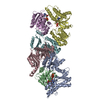
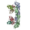
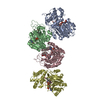
| Title | Solution Structure of the Enzyme I dimer Using Residual Dipolar Couplings and Small Angle X-Ray Scattering | ||||||
 Components Components | Phosphoenolpyruvate-protein phosphotransferase Phosphoenolpyruvate—protein phosphotransferase Phosphoenolpyruvate—protein phosphotransferase | ||||||
 Keywords Keywords | TRANSFERASE / SUGAR PHOSPHOTRANSFERASE SYSTEM / PTS | ||||||
| Function / homology |  Function and homology informationphosphoenolpyruvate-protein phosphotransferase / phosphoenolpyruvate-protein phosphotransferase activity / phosphoenolpyruvate-dependent sugar phosphotransferase system / kinase activity / phosphorylation / identical protein binding / metal ion binding / cytosol Function and homology informationphosphoenolpyruvate-protein phosphotransferase / phosphoenolpyruvate-protein phosphotransferase activity / phosphoenolpyruvate-dependent sugar phosphotransferase system / kinase activity / phosphorylation / identical protein binding / metal ion binding / cytosolSimilarity search - Function | ||||||
| Biological species |  Escherichia coli (E. coli) Escherichia coli (E. coli) | ||||||
| Method | SOLUTION NMR / SOLUTION SCATTERING / simulated annealing | ||||||
| Model details | regularized mean of the 99 structures that are reported in primary citation | ||||||
 Authors Authors | Schwieters, C.D. / Suh, J. / Grishaev, A. / Takayama, Y. / Guirlando, R. / Clore, G. | ||||||
 Citation Citation | Journal: J Am Chem Soc / Year: 2010 Title: Solution structure of the 128 kDa enzyme I dimer from Escherichia coli and its 146 kDa complex with HPr using residual dipolar couplings and small- and wide-angle X-ray scattering. Authors: Charles D Schwieters / Jeong-Yong Suh / Alexander Grishaev / Rodolfo Ghirlando / Yuki Takayama / G Marius Clore /  Abstract: The solution structures of free Enzyme I (EI, ∼128 kDa, 575 × 2 residues), the first enzyme in the bacterial phosphotransferase system, and its complex with HPr (∼146 kDa) have been solved using ...The solution structures of free Enzyme I (EI, ∼128 kDa, 575 × 2 residues), the first enzyme in the bacterial phosphotransferase system, and its complex with HPr (∼146 kDa) have been solved using novel methodology that makes use of prior structural knowledge (namely, the structures of the dimeric EIC domain and the isolated EIN domain both free and complexed to HPr), combined with residual dipolar coupling (RDC), small- (SAXS) and wide- (WAXS) angle X-ray scattering and small-angle neutron scattering (SANS) data. The calculational strategy employs conjoined rigid body/torsion/Cartesian simulated annealing, and incorporates improvements in calculating and refining against SAXS/WAXS data that take into account complex molecular shapes in the description of the solvent layer resulting in a better representation of the SAXS/WAXS data. The RDC data orient the symmetrically related EIN domains relative to the C(2) symmetry axis of the EIC dimer, while translational, shape, and size information is provided by SAXS/WAXS. The resulting structures are independently validated by SANS. Comparison of the structures of the free EI and the EI-HPr complex with that of the crystal structure of a trapped phosphorylated EI intermediate reveals large (∼70-90°) hinge body rotations of the two subdomains comprising the EIN domain, as well as of the EIN domain relative to the dimeric EIC domain. These large-scale interdomain motions shed light on the structural transitions that accompany the catalytic cycle of EI. | ||||||
| History |
|
- Structure visualization
| Structure viewer | Molecule: MolmilJmol/JSmol |
|---|
- Downloads & links
-Download
| PDBx/mmCIF format | 2kx9.cif.gz | 758.8 KB | Display | PDBx/mmCIF format |
|---|---|---|---|---|
| PDB format | pdb2kx9.ent.gz | 639.3 KB | Display | PDB format |
| PDBx/mmJSON format | 2kx9.json.gz | Tree view | PDBx/mmJSON format | |
| Others |  Other downloads Other downloads |
-Validation report
| Arichive directory | https://data.pdbj.org/pub/pdb/validation_reports/kx/2kx9ftp://data.pdbj.org/pub/pdb/validation_reports/kx/2kx9 | HTTPS FTP |
|---|
-Related structure data
| Related structure data | 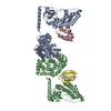 2xdfC C: citing same article ( |
|---|---|
| Similar structure data | |
| Other databases |
|


-Links
 PDBj
PDBj
- Assembly
| Deposited unit | 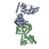
| |||||||||
|---|---|---|---|---|---|---|---|---|---|---|
| 1 |
| |||||||||
| NMR ensembles |
|
-Components
| #1: Protein | Phosphoenolpyruvate—protein phosphotransferase / Phosphotransferase system / enzyme I Mass: 63419.344 Da / Num. of mol.: 2 / Fragment: UNP residues 1-573 Source method: isolated from a genetically manipulated source Source: (gene. exp.) Escherichia coli (E. coli) / Strain: K12 / Gene: ptsI, b2416, JW2409 / Production host: Escherichia coli (E. coli) / Strain (production host): BL21 StarTM (DE3)References: UniProt: P08839, phosphoenolpyruvate-protein phosphotransferase |
|---|
-Experimental details
-Experiment
| Experiment |
| |||
|---|---|---|---|---|
| NMR experiment | Type: TROSY-based 1H-15N correlation spectroscopy |
- Sample preparation
| Details | Contents: 20 mM TRIS, 100 mM sodium chloride, 10 mM DTT, 4 mM MgCl2, 1 mM EDTA, 10 % D2O, 1 tablet protease inhibitor, 0.15 mM EI dimer, 90% H2O/10% D2O Solvent system: 90% H2O/10% D2O | |||||||||||||||||||||||||||
|---|---|---|---|---|---|---|---|---|---|---|---|---|---|---|---|---|---|---|---|---|---|---|---|---|---|---|---|---|
| Sample |
| |||||||||||||||||||||||||||
| Sample conditions | pH: 7.4 / Pressure: ambient / Temperature: 310 K |
-Data collection
| NMR spectrometer | Type: Bruker DRX / Manufacturer: Bruker / Model: DRX / Field strength: 800 MHz | |||||||||||||||||||||||||||
|---|---|---|---|---|---|---|---|---|---|---|---|---|---|---|---|---|---|---|---|---|---|---|---|---|---|---|---|---|
| Soln scatter | Data analysis software list: GNOM / Protein length: 150 / Sample pH: 7.4 / Temperature: 298 K
|
- Processing
| NMR software |
| ||||||||||||
|---|---|---|---|---|---|---|---|---|---|---|---|---|---|
| Refinement | Method: simulated annealing / Software ordinal: 1 Details: STRUCTURE STATISTICS: MODEL 1: SAXS CHI2 Q->0.44: 0.43 SAXS CHI2 FULL RANGE: 2.26 SANS CHI2: 0.39 RDC R-FACTOR: 18.03 % RDC DA: 14.5 HZ RDC RH: 0.49 MODEL 2: SAXS CHI2 Q->0.44: 0.40 SAXS ...Details: STRUCTURE STATISTICS: MODEL 1: SAXS CHI2 Q->0.44: 0.43 SAXS CHI2 FULL RANGE: 2.26 SANS CHI2: 0.39 RDC R-FACTOR: 18.03 % RDC DA: 14.5 HZ RDC RH: 0.49 MODEL 2: SAXS CHI2 Q->0.44: 0.40 SAXS CHI2 FULL RANGE: 1.99 SANS CHI2: 0.58 RDC R-FACTOR: 18.07 RDC DA: 14.5 HZ RDC RH: 0.48 AVERAGE OVER THE FULL 99-MEMBER ENSEMBLE: SAXS CHI2 Q->0.44: 0.29 +/- 0.05 SAXS CHI2 FULL RANGE: 1.34 +/- 0.27 SANS CHI2: 0.41 +/- 0.08 RDC R-FACTOR: 18.07 +/- 0.02 % RDC DA: 14.5 +/- 0.1 HZ RDC RH: 0.49 +/- 0.00 | ||||||||||||
| NMR representative | Selection criteria: closest to the average | ||||||||||||
| NMR ensemble | Conformer selection criteria: target function / Conformers calculated total number: 120 / Conformers submitted total number: 2 | ||||||||||||
| Soln scatter model | Conformer selection criteria: REGULARIZED MEAN OF 99 MODELS Details: The initial structure of the EI dimer was constructed as a hybrid of the crystal structure of phosphorylated EI intermediate captured by the inhibitor oxalate (PDB code 2HWG) and the NMR ...Details: The initial structure of the EI dimer was constructed as a hybrid of the crystal structure of phosphorylated EI intermediate captured by the inhibitor oxalate (PDB code 2HWG) and the NMR structure of the EIN-HPr complex (PDB code 3EZA). Throughout the structure determination, the backbone atomic coordinates of each EIN domain (residues 1-254) were treated as rigid bodies, with the two symmetry related EIC domains (residues 262- 573) held fixed in space. Coordinates in the linker region (residues 255-261) were allowed varying degrees of freedom during the calculation through the use of the internal variable module (IVM) of Xplor-NIH. This entry corresponds to the regularized mean of the 99 structures for which data was reported in the primary publication, with the B-factor column representing the per-atom spread (in B-factor units). The calculated structural statistics for the original 99 structures and for the regularized mean are shown below. Entry fitting list: PDB ENTRIES 2HWG AND 3EZA / Num. of conformers calculated: 99 / Num. of conformers submitted: 1 / Representative conformer: 1 / Software list: GNOM,XPLOR-NIH |
