 Movie
Movie Controller


+ Open data

- Basic information
| Entry | Database: PDB / ID: 2jac | ||||||
|---|---|---|---|---|---|---|---|
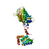
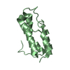
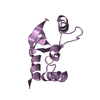
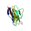
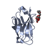
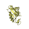
| Title | Glutaredoxin Grx1p C30S mutant from yeast | ||||||
 Components Components | GLUTAREDOXIN-1 GLRX GLRX | ||||||
 Keywords Keywords | ELECTRON TRANSPORT / REDOX-ACTIVE CENTER / OXIDOREDUCTASE / GLUTATHIONE / GLUTAREDOXIN | ||||||
| Function / homology |  Function and homology informationprotein glutathionylation / glutathione peroxidase / glutathione disulfide oxidoreductase activity / glutathione peroxidase activity / glutathione transferase / glutathione transferase activity / cellular response to oxidative stress / nucleus / cytoplasm Function and homology informationprotein glutathionylation / glutathione peroxidase / glutathione disulfide oxidoreductase activity / glutathione peroxidase activity / glutathione transferase / glutathione transferase activity / cellular response to oxidative stress / nucleus / cytoplasmSimilarity search - Function | ||||||
| Biological species |  SACCHAROMYCES CEREVISIAE (brewer's yeast) SACCHAROMYCES CEREVISIAE (brewer's yeast) | ||||||
| Method | X-RAY DIFFRACTION / SYNCHROTRON / MOLECULAR REPLACEMENT / Resolution: 2.02 Å | ||||||
 Authors Authors | Hakansson, K.O. / Winther, J.R. | ||||||
 Citation Citation | Journal: Acta Crystallogr.,Sect.D / Year: 2007 Title: Structure of Glutaredoxin Grx1P C30S Mutant from Yeast. Authors: Hakansson, K.O. / Winther, J.R. #1: Journal: Acta Crystallogr.,Sect.F / Year: 2006 Title: Crystallisation of Mutant Forms of Glutaredoxin Grx1P from Yeast Authors: Hakansson, K.O. / Ostergaard, H. / Winther, J.R. | ||||||
| History |
|
- Structure visualization
| Structure viewer | Molecule: MolmilJmol/JSmol |
|---|
- Downloads & links
-Download
| PDBx/mmCIF format | 2jac.cif.gz | 34.9 KB | Display | PDBx/mmCIF format |
|---|---|---|---|---|
| PDB format | pdb2jac.ent.gz | 23.7 KB | Display | PDB format |
| PDBx/mmJSON format | 2jac.json.gz | Tree view | PDBx/mmJSON format | |
| Others |  Other downloads Other downloads |
-Validation report
| Arichive directory | https://data.pdbj.org/pub/pdb/validation_reports/ja/2jacftp://data.pdbj.org/pub/pdb/validation_reports/ja/2jac | HTTPS FTP |
|---|
-Related structure data


-Links
 PDBj
PDBj


- Assembly
| Deposited unit | 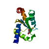
| ||||||||
|---|---|---|---|---|---|---|---|---|---|
| 1 |
| ||||||||
| Unit cell |
| ||||||||
| Components on special symmetry positions |
|
-Components
| #1: Protein | GLRX / GLUTAREDOXIN / GLUTATHIONE-DEPENDENT OXIDOREDUCTASE 1 Mass: 12378.087 Da / Num. of mol.: 1 / Mutation: YES Source method: isolated from a genetically manipulated source Source: (gene. exp.) SACCHAROMYCES CEREVISIAE (brewer's yeast)Production host:  ESCHERICHIA COLI (E. coli) / Strain (production host): BL21(DE3) / References: UniProt: P25373 ESCHERICHIA COLI (E. coli) / Strain (production host): BL21(DE3) / References: UniProt: P25373 |
|---|---|
| #2: Chemical | ChemComp-GSH / Glutathione  Mass: 307.323 Da / Num. of mol.: 1 / Source method: obtained synthetically / Formula: C10H17N3O6S Mass: 307.323 Da / Num. of mol.: 1 / Source method: obtained synthetically / Formula: C10H17N3O6S |
| #3: Water | ChemComp-HOH / Water Mass: 18.015 Da / Num. of mol.: 65 / Source method: isolated from a natural source / Formula: H2O Mass: 18.015 Da / Num. of mol.: 65 / Source method: isolated from a natural source / Formula: H2O |
| Compound details | ENGINEERED |
-Experimental details
-Experiment
| Experiment | Method: X-RAY DIFFRACTION / Number of used crystals: 1 |
|---|
- Sample preparation
| Crystal | Density Matthews: 2.5 Å3/Da / Density % sol: 52 % |
|---|---|
| Crystal grow | pH: 4.75 / Details: 50MM CITRATE PH 4.75 10-22.5% PEG 4000 |
-Data collection
| Diffraction | Mean temperature: 100 K |
|---|---|
| Diffraction source | Source: SYNCHROTRON / Site: MAX II  / Beamline: I711 / Wavelength: 1.115 / Beamline: I711 / Wavelength: 1.115 |
| Detector | Type: MARRESEARCH / Detector: CCD / Date: Oct 30, 2005 |
| Radiation | Protocol: SINGLE WAVELENGTH / Monochromatic (M) / Laue (L): M / Scattering type: x-ray |
| Radiation wavelength | Wavelength: 1.115 Å / Relative weight: 1 |
| Reflection | Resolution: 2.02→20 Å / Num. obs: 8364 / % possible obs: 97.5 % / Observed criterion σ(I): -10 / Redundancy: 4.1 % / Rmerge(I) obs: 0.06 / Net I/σ(I): 8.9 |
| Reflection shell | Resolution: 2.02→2.13 Å / Redundancy: 4 % / Rmerge(I) obs: 0.23 / Mean I/σ(I) obs: 3.1 / % possible all: 84.5 |
- Processing
| Software |
| ||||||||||||||||||||||||||||||||||||||||||||||||||||||||||||
|---|---|---|---|---|---|---|---|---|---|---|---|---|---|---|---|---|---|---|---|---|---|---|---|---|---|---|---|---|---|---|---|---|---|---|---|---|---|---|---|---|---|---|---|---|---|---|---|---|---|---|---|---|---|---|---|---|---|---|---|---|---|
| Refinement | Method to determine structure: MOLECULAR REPLACEMENT / Resolution: 2.02→20 Å / Cross valid method: THROUGHOUT / σ(F): 0
| ||||||||||||||||||||||||||||||||||||||||||||||||||||||||||||
| Refinement step | Cycle: LAST / Resolution: 2.02→20 Å
| ||||||||||||||||||||||||||||||||||||||||||||||||||||||||||||
| Refine LS restraints |
|
