 Movie
Movie Controller


+ Open data

- Basic information
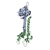
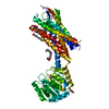
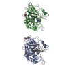
| Entry | Database: PDB / ID: 2jad | ||||||
|---|---|---|---|---|---|---|---|
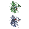
| Title | Yellow fluorescent protein - glutaredoxin fusion protein | ||||||
 Components Components | YELLOW FLUORESCENT PROTEIN GLUTAREDOXIN FUSION PROTEIN | ||||||
 Keywords Keywords |  ELECTRON TRANSPORT / YELLOW FLUORESCENT PROTEIN / REDOX- ACTIVE CENTER / YEAST / GRX1P / TRANSPORT / GLUTAREDOXIN ELECTRON TRANSPORT / YELLOW FLUORESCENT PROTEIN / REDOX- ACTIVE CENTER / YEAST / GRX1P / TRANSPORT / GLUTAREDOXIN | ||||||
| Function / homology |  Function and homology informationprotein glutathionylation / glutathione peroxidase / glutathione disulfide oxidoreductase activity / glutathione peroxidase activity / glutathione transferase / glutathione transferase activity / bioluminescence / generation of precursor metabolites and energy / cellular response to oxidative stress / nucleus / cytoplasm Function and homology informationprotein glutathionylation / glutathione peroxidase / glutathione disulfide oxidoreductase activity / glutathione peroxidase activity / glutathione transferase / glutathione transferase activity / bioluminescence / generation of precursor metabolites and energy / cellular response to oxidative stress / nucleus / cytoplasmSimilarity search - Function | ||||||
| Biological species |   AEQUOREA VICTORIA (jellyfish) AEQUOREA VICTORIA (jellyfish) SACCHAROMYCES CEREVISIAE (brewer's yeast) SACCHAROMYCES CEREVISIAE (brewer's yeast) | ||||||
| Method | X-RAY DIFFRACTION / SYNCHROTRON / MOLECULAR REPLACEMENT / Resolution: 2.7 Å | ||||||
 Authors Authors | Hakansson, K.O. / Winther, J.R. | ||||||
 Citation Citation | Journal: Acta Crystallogr.,Sect.D / Year: 2007 Title: Structure of Glutaredoxin Grx1P C30S Mutant from Yeast. Authors: Hakansson, K.O. / Winther, J.R. #1: Journal: Acta Crystallogr.,Sect.F / Year: 2006 Title: Crystallisation of Mutant Forms of Glutaredoxin Grx1P from Yeast Authors: Hakansson, K.O. / Winther, J.R. | ||||||
| History |
| ||||||
| Remark 700 | SHEET DETERMINATION METHOD: DSSP THE SHEETS PRESENTED AS "AA" IN EACH CHAIN ON SHEET RECORDS BELOW ... SHEET DETERMINATION METHOD: DSSP THE SHEETS PRESENTED AS "AA" IN EACH CHAIN ON SHEET RECORDS BELOW IS ACTUALLY AN 12-STRANDED BARREL THIS IS REPRESENTED BY A 13-STRANDED SHEET IN WHICH THE FIRST AND LAST STRANDS ARE IDENTICAL. |
- Structure visualization
| Structure viewer | Molecule: MolmilJmol/JSmol |
|---|
- Downloads & links
-Download
| PDBx/mmCIF format | 2jad.cif.gz | 78.2 KB | Display | PDBx/mmCIF format |
|---|---|---|---|---|
| PDB format | pdb2jad.ent.gz | 58.2 KB | Display | PDB format |
| PDBx/mmJSON format | 2jad.json.gz | Tree view | PDBx/mmJSON format | |
| Others |  Other downloads Other downloads |
-Validation report
| Arichive directory | https://data.pdbj.org/pub/pdb/validation_reports/ja/2jadftp://data.pdbj.org/pub/pdb/validation_reports/ja/2jad | HTTPS FTP |
|---|
-Related structure data
| Related structure data |  2jacC 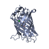 1h6rS S: Starting model for refinement C: citing same article ( |
|---|---|
| Similar structure data |


-Links
 PDBj
PDBj


- Assembly
| Deposited unit | 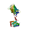
| ||||||||
|---|---|---|---|---|---|---|---|---|---|
| 1 |
| ||||||||
| Unit cell |
|
-Components
| #1: Protein | Mass: 40975.215 Da / Num. of mol.: 1 / Mutation: YES Source method: isolated from a genetically manipulated source Details: THE MUTATED RESIDUE NUMBER 276 CORRESPONDS TO C30S IN GLUTAREDOXIN Source: (gene. exp.) AEQUOREA VICTORIA (jellyfish), (gene. exp.) SACCHAROMYCES CEREVISIAE (brewer's yeast)Production host:  ESCHERICHIA COLI (E. coli) / Strain (production host): BL21(DE3) / References: UniProt: P42212, UniProt: P25373 ESCHERICHIA COLI (E. coli) / Strain (production host): BL21(DE3) / References: UniProt: P42212, UniProt: P25373 | ||
|---|---|---|---|
| #2: Chemical | Sulfate  Mass: 96.063 Da / Num. of mol.: 2 / Source method: obtained synthetically / Formula: SO4 Mass: 96.063 Da / Num. of mol.: 2 / Source method: obtained synthetically / Formula: SO4#3: Water | ChemComp-HOH / | Water Mass: 18.015 Da / Num. of mol.: 55 / Source method: isolated from a natural source / Formula: H2O Mass: 18.015 Da / Num. of mol.: 55 / Source method: isolated from a natural source / Formula: H2O |
-Experimental details
-Experiment
| Experiment | Method: X-RAY DIFFRACTION / Number of used crystals: 1 |
|---|
- Sample preparation
| Crystal | Density Matthews: 3.6 Å3/Da / Density % sol: 66 % |
|---|---|
| Crystal grow | pH: 10 / Details: 50MM BICARBONATE PH 10 1.5-1.75M MGSO4 |
-Data collection
| Diffraction | Mean temperature: 100 K |
|---|---|
| Diffraction source | Source: SYNCHROTRON / Site: MAX II  / Beamline: I711 / Wavelength: 1.115 / Beamline: I711 / Wavelength: 1.115 |
| Detector | Type: MARRESEARCH / Detector: CCD / Date: Oct 30, 2005 |
| Radiation | Protocol: SINGLE WAVELENGTH / Monochromatic (M) / Laue (L): M / Scattering type: x-ray |
| Radiation wavelength | Wavelength: 1.115 Å / Relative weight: 1 |
| Reflection | Resolution: 2.7→20 Å / Num. obs: 15714 / % possible obs: 96.7 % / Observed criterion σ(I): -10 / Redundancy: 2.8 % / Rmerge(I) obs: 0.1 / Net I/σ(I): 5.8 |
| Reflection shell | Resolution: 2.7→2.85 Å / Redundancy: 2.4 % / Rmerge(I) obs: 0.36 / Mean I/σ(I) obs: 2.1 / % possible all: 95.1 |
- Processing
| Software |
| ||||||||||||||||||||||||||||||||||||||||||||||||||||||||||||
|---|---|---|---|---|---|---|---|---|---|---|---|---|---|---|---|---|---|---|---|---|---|---|---|---|---|---|---|---|---|---|---|---|---|---|---|---|---|---|---|---|---|---|---|---|---|---|---|---|---|---|---|---|---|---|---|---|---|---|---|---|---|
| Refinement | Method to determine structure: MOLECULAR REPLACEMENT Starting model: PDB ENTRY 1H6R Resolution: 2.7→20 Å / Cross valid method: THROUGHOUT / σ(F): 0 Details: RESIDUES 237-241 BUILT AS AAAGG AND RESIDUES 242-245 ABSENT DUE TO DISORDER. THE C-TERMINUS WAS ALSO DISORDERED
| ||||||||||||||||||||||||||||||||||||||||||||||||||||||||||||
| Refinement step | Cycle: LAST / Resolution: 2.7→20 Å
| ||||||||||||||||||||||||||||||||||||||||||||||||||||||||||||
| Refine LS restraints |
|
