 Movie
Movie Controller

[English] 日本語
 Yorodumi
- PDB-3rhb: Crystal structure of the apo form of glutaredoxin C5 from Arabido... -
+ Open data

- Basic information
| Entry | Database: PDB / ID: 3rhb | ||||||
|---|---|---|---|---|---|---|---|
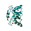
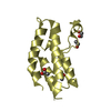
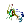
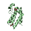
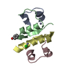
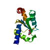
| Title | Crystal structure of the apo form of glutaredoxin C5 from Arabidopsis thaliana | ||||||
 Components Components | Glutaredoxin-C5, chloroplastic | ||||||
 Keywords Keywords |  OXIDOREDUCTASE / Thioredoxin fold / thiol-disulfide oxidoreductase / glutaredoxin OXIDOREDUCTASE / Thioredoxin fold / thiol-disulfide oxidoreductase / glutaredoxin | ||||||
| Function / homology |  Function and homology information Function and homology information | ||||||
| Biological species |  Arabidopsis thaliana (thale cress) Arabidopsis thaliana (thale cress) | ||||||
| Method | X-RAY DIFFRACTION / SYNCHROTRON / MOLECULAR REPLACEMENT / Resolution: 1.2 Å | ||||||
 Authors Authors | Roret, T. / Couturier, J. / Tsan, P. / Jacquot, J.P. / Rouhier, N. / Didierjean, C. | ||||||
 Citation Citation | Journal: J.Biol.Chem. / Year: 2011 Title: Arabidopsis chloroplastic glutaredoxin c5 as a model to explore molecular determinants for iron-sulfur cluster binding into glutaredoxins. Authors: Couturier, J. / Stroher, E. / Albetel, A.N. / Roret, T. / Muthuramalingam, M. / Tarrago, L. / Seidel, T. / Tsan, P. / Jacquot, J.P. / Johnson, M.K. / Dietz, K.J. / Didierjean, C. / Rouhier, N. | ||||||
| History |
|
- Structure visualization
| Structure viewer | Molecule: MolmilJmol/JSmol |
|---|
- Downloads & links
-Download
| PDBx/mmCIF format | 3rhb.cif.gz | 58.9 KB | Display | PDBx/mmCIF format |
|---|---|---|---|---|
| PDB format | pdb3rhb.ent.gz | 42.4 KB | Display | PDB format |
| PDBx/mmJSON format | 3rhb.json.gz | Tree view | PDBx/mmJSON format | |
| Others |  Other downloads Other downloads |
-Validation report
| Arichive directory | https://data.pdbj.org/pub/pdb/validation_reports/rh/3rhbftp://data.pdbj.org/pub/pdb/validation_reports/rh/3rhb | HTTPS FTP |
|---|
-Related structure data
| Related structure data | 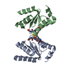 3rhcC 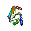 3fz9S S: Starting model for refinement C: citing same article ( |
|---|---|
| Similar structure data |


-Links
 PDBj
PDBj

- Assembly
| Deposited unit | 
| |||||||||
|---|---|---|---|---|---|---|---|---|---|---|
| 1 |
| |||||||||
| Unit cell |
| |||||||||
| Components on special symmetry positions |
| |||||||||
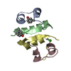
| Details | AUTHORS STATE THAT THE PROTEIN IS MONOMERIC IN SOLUTION ACCORDING TO ANALYTICAL GEL FILTRATION ANALYSES. |
-Components
| #1: Protein | Mass: 12498.430 Da / Num. of mol.: 1 Source method: isolated from a genetically manipulated source Source: (gene. exp.) Arabidopsis thaliana (thale cress) / Gene: GRXC5, At4g28730, F16A16.160 / Production host:  Escherichia coli (E. coli) / References: UniProt: Q8GWS0 Escherichia coli (E. coli) / References: UniProt: Q8GWS0 | ||
|---|---|---|---|
| #2: Chemical | ChemComp-GSH / Glutathione  Mass: 307.323 Da / Num. of mol.: 1 / Source method: obtained synthetically / Formula: C10H17N3O6S Mass: 307.323 Da / Num. of mol.: 1 / Source method: obtained synthetically / Formula: C10H17N3O6S | ||
| #3: Chemical | Sulfate  Mass: 96.063 Da / Num. of mol.: 3 / Source method: obtained synthetically / Formula: SO4 Mass: 96.063 Da / Num. of mol.: 3 / Source method: obtained synthetically / Formula: SO4#4: Water | ChemComp-HOH / | Water Mass: 18.015 Da / Num. of mol.: 108 / Source method: isolated from a natural source / Formula: H2O Mass: 18.015 Da / Num. of mol.: 108 / Source method: isolated from a natural source / Formula: H2O |
-Experimental details
-Experiment
| Experiment | Method: X-RAY DIFFRACTION / Number of used crystals: 1 |
|---|
- Sample preparation
| Crystal | Density Matthews: 1.86 Å3/Da / Density % sol: 34.03 % |
|---|---|
| Crystal grow | Temperature: 277 K / Method: microbatch Details: 1.6 M ammonium sulfate, 500 mM lithium chloride, Microbatch, temperature 277K |
-Data collection
| Diffraction | Mean temperature: 100 K |
|---|---|
| Diffraction source | Source: SYNCHROTRON / Site: ESRF  / Beamline: BM30A / Wavelength: 0.97926 Å / Beamline: BM30A / Wavelength: 0.97926 Å |
| Detector | Type: ADSC QUANTUM 315r / Detector: CCD / Date: May 25, 2010 / Details: mirrors |
| Radiation | Monochromator: 111 SILICON SINGLE CRYSTAL / Protocol: SINGLE WAVELENGTH / Monochromatic (M) / Laue (L): M / Scattering type: x-ray |
| Radiation wavelength | Wavelength: 0.97926 Å / Relative weight: 1 |
| Reflection | Resolution: 1.2→33.25 Å / Num. obs: 26715 / % possible obs: 94 % / Observed criterion σ(F): 0 / Observed criterion σ(I): 0 / Redundancy: 7.5 % / Rmerge(I) obs: 0.052 / Net I/σ(I): 21.9 |
- Processing
| Software |
| ||||||||||||||||||||||||||||||||||||||||||||||||||||||||||||||||||||||
|---|---|---|---|---|---|---|---|---|---|---|---|---|---|---|---|---|---|---|---|---|---|---|---|---|---|---|---|---|---|---|---|---|---|---|---|---|---|---|---|---|---|---|---|---|---|---|---|---|---|---|---|---|---|---|---|---|---|---|---|---|---|---|---|---|---|---|---|---|---|---|---|
| Refinement | Method to determine structure: MOLECULAR REPLACEMENT Starting model: PDB ENTRY 3FZ9 Resolution: 1.2→33.25 Å / Cor.coef. Fo:Fc: 0.968 / Cor.coef. Fo:Fc free: 0.961 / SU B: 1.015 / SU ML: 0.022 / Cross valid method: THROUGHOUT / ESU R Free: 0.042 / Stereochemistry target values: MAXIMUM LIKELIHOOD
| ||||||||||||||||||||||||||||||||||||||||||||||||||||||||||||||||||||||
| Solvent computation | Ion probe radii: 0.8 Å / Shrinkage radii: 0.8 Å / VDW probe radii: 1.4 Å / Solvent model: MASK | ||||||||||||||||||||||||||||||||||||||||||||||||||||||||||||||||||||||
| Displacement parameters | Biso mean: 18.551 Å2
| ||||||||||||||||||||||||||||||||||||||||||||||||||||||||||||||||||||||
| Refinement step | Cycle: LAST / Resolution: 1.2→33.25 Å
| ||||||||||||||||||||||||||||||||||||||||||||||||||||||||||||||||||||||
| Refine LS restraints |
| ||||||||||||||||||||||||||||||||||||||||||||||||||||||||||||||||||||||
| LS refinement shell | Resolution: 1.2→1.231 Å / Total num. of bins used: 20
|
