 Movie
Movie Controller


+ Open data

- Basic information
| Entry | Database: PDB / ID: 2j2j | ||||||
|---|---|---|---|---|---|---|---|
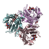
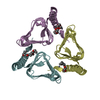
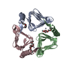
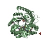
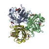
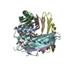
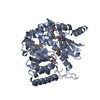
| Title | Canine adenovirus fibre head at 1.5 A resolution | ||||||
 Components Components | FIBER PROTEIN Fibrous protein Fibrous protein | ||||||
 Keywords Keywords | VIRAL PROTEIN / FIBER PROTEIN / CANINE ADENOVIRUS / AD / CAR / KNOB / HEAD / FIBER FIBRE / ADENOVIRUS / COXSACKIEVIRUS / ADENOVIRUS RECEPTOR | ||||||
| Function / homology |  Function and homology information Function and homology informationadhesion receptor-mediated virion attachment to host cell / viral capsid / cell adhesion / symbiont entry into host cell / host cell nucleusSimilarity search - Function | ||||||
| Biological species |  CANINE ADENOVIRUS 2 CANINE ADENOVIRUS 2 | ||||||
| Method | X-RAY DIFFRACTION / SYNCHROTRON / MOLECULAR REPLACEMENT / Resolution: 1.5 Å | ||||||
 Authors Authors | Seiradake, E. / Lortat-Jacob, H. / Billet, O. / Kremer, E.J. / Cusack, S. | ||||||
 Citation Citation | Journal: J.Biol.Chem. / Year: 2006 Title: Structural and Mutational Analysis of Human Ad37 and Canine Adenovirus 2 Fiber Heads in Complex with the D1 Domain of Coxsackie and Adenovirus Receptor. Authors: Seiradake, E. / Lortat-Jacob, H. / Billet, O. / Kremer, E.J. / Cusack, S. | ||||||
| History |
| ||||||
| Remark 700 | SHEET THE SHEET STRUCTURE OF THIS MOLECULE IS BIFURCATED. IN ORDER TO REPRESENT THIS FEATURE IN ... SHEET THE SHEET STRUCTURE OF THIS MOLECULE IS BIFURCATED. IN ORDER TO REPRESENT THIS FEATURE IN THE SHEET RECORDS BELOW, TWO SHEETS ARE DEFINED. |
- Structure visualization
| Structure viewer | Molecule: MolmilJmol/JSmol |
|---|
- Downloads & links
-Download
| PDBx/mmCIF format | 2j2j.cif.gz | 467.7 KB | Display | PDBx/mmCIF format |
|---|---|---|---|---|
| PDB format | pdb2j2j.ent.gz | 382.9 KB | Display | PDB format |
| PDBx/mmJSON format | 2j2j.json.gz | Tree view | PDBx/mmJSON format | |
| Others |  Other downloads Other downloads |
-Validation report
| Arichive directory | https://data.pdbj.org/pub/pdb/validation_reports/j2/2j2jftp://data.pdbj.org/pub/pdb/validation_reports/j2/2j2j | HTTPS FTP |
|---|
-Related structure data
| Related structure data |  2j12C  2j1kC 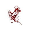 1qhvS C: citing same article ( S: Starting model for refinement |
|---|---|
| Similar structure data |

-Links
 PDBj
PDBj
- Assembly
| Deposited unit | 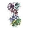
| ||||||||||||||||||||||||
|---|---|---|---|---|---|---|---|---|---|---|---|---|---|---|---|---|---|---|---|---|---|---|---|---|---|
| 1 | 
| ||||||||||||||||||||||||
| 2 | 
| ||||||||||||||||||||||||
| Unit cell |
| ||||||||||||||||||||||||
| Noncrystallographic symmetry (NCS) | NCS oper:
|
-Components
| #1: Protein | Fibrous protein / PIV / CANINE ADENOVIRUS 2 FIBRE HEAD Mass: 21685.541 Da / Num. of mol.: 6 / Fragment: FIBRE HEAD DOMAIN, RESIDUES 358-542 Source method: isolated from a genetically manipulated source Source: (gene. exp.) CANINE ADENOVIRUS 2 / Plasmid: PQE / Production host:  ESCHERICHIA COLI (E. coli) / Strain (production host): M15 / References: UniProt: Q65914 ESCHERICHIA COLI (E. coli) / Strain (production host): M15 / References: UniProt: Q65914#2: Water | ChemComp-HOH / | Water Mass: 18.015 Da / Num. of mol.: 1539 / Source method: isolated from a natural source / Formula: H2O Mass: 18.015 Da / Num. of mol.: 1539 / Source method: isolated from a natural source / Formula: H2OSequence details | CONTAINS ONLY FIBRE HEAD DOMAIN, AND N-TERMINAL 6XHIS TAG | |
|---|
-Experimental details
-Experiment
| Experiment | Method: X-RAY DIFFRACTION / Number of used crystals: 1 |
|---|
- Sample preparation
| Crystal | Density Matthews: 2.2 Å3/Da / Density % sol: 44 % |
|---|---|
| Crystal grow | Details: 0.15 M KBR, 30% PEG MONOMETHYL-ETHER 2000 |
-Data collection
| Diffraction | Mean temperature: 100 K |
|---|---|
| Diffraction source | Source: SYNCHROTRON / Site: ESRF  / Beamline: ID14-1 / Wavelength: 0.934 / Beamline: ID14-1 / Wavelength: 0.934 |
| Detector | Type: ADSC CCD / Detector: CCD |
| Radiation | Protocol: SINGLE WAVELENGTH / Monochromatic (M) / Laue (L): M / Scattering type: x-ray |
| Radiation wavelength | Wavelength: 0.934 Å / Relative weight: 1 |
| Reflection | Resolution: 1.5→28.4 Å / Num. obs: 156532 / % possible obs: 95.1 % / Observed criterion σ(I): 0 / Redundancy: 4.1 % / Biso Wilson estimate: 8.1 Å2 / Rmerge(I) obs: 0.1 / Net I/σ(I): 15.3 |
| Reflection shell | Resolution: 1.5→1.54 Å / Redundancy: 3.8 % / Rmerge(I) obs: 0.15 / Mean I/σ(I) obs: 7.8 / % possible all: 89.4 |
- Processing
| Software |
| ||||||||||||||||||||||||||||||||||||||||||||||||||||||||||||||||||||||||||||||||||||||||||||||||||||||||||||||||||||||||||||||||||||||||||||||||||||||||||||||||||||||||||||||||||||||
|---|---|---|---|---|---|---|---|---|---|---|---|---|---|---|---|---|---|---|---|---|---|---|---|---|---|---|---|---|---|---|---|---|---|---|---|---|---|---|---|---|---|---|---|---|---|---|---|---|---|---|---|---|---|---|---|---|---|---|---|---|---|---|---|---|---|---|---|---|---|---|---|---|---|---|---|---|---|---|---|---|---|---|---|---|---|---|---|---|---|---|---|---|---|---|---|---|---|---|---|---|---|---|---|---|---|---|---|---|---|---|---|---|---|---|---|---|---|---|---|---|---|---|---|---|---|---|---|---|---|---|---|---|---|---|---|---|---|---|---|---|---|---|---|---|---|---|---|---|---|---|---|---|---|---|---|---|---|---|---|---|---|---|---|---|---|---|---|---|---|---|---|---|---|---|---|---|---|---|---|---|---|---|---|
| Refinement | Method to determine structure: MOLECULAR REPLACEMENT Starting model: PDB ENTRY 1QHV Resolution: 1.5→106 Å / Cor.coef. Fo:Fc: 0.946 / Cor.coef. Fo:Fc free: 0.918 / SU B: 2.675 / SU ML: 0.047 / Cross valid method: THROUGHOUT / ESU R: 0.101 / ESU R Free: 0.081 / Stereochemistry target values: MAXIMUM LIKELIHOOD / Details: HYDROGENS HAVE BEEN ADDED IN THE RIDING POSITIONS
| ||||||||||||||||||||||||||||||||||||||||||||||||||||||||||||||||||||||||||||||||||||||||||||||||||||||||||||||||||||||||||||||||||||||||||||||||||||||||||||||||||||||||||||||||||||||
| Solvent computation | Ion probe radii: 0.8 Å / Shrinkage radii: 0.8 Å / VDW probe radii: 1.4 Å / Solvent model: MASK | ||||||||||||||||||||||||||||||||||||||||||||||||||||||||||||||||||||||||||||||||||||||||||||||||||||||||||||||||||||||||||||||||||||||||||||||||||||||||||||||||||||||||||||||||||||||
| Displacement parameters | Biso mean: 6.01 Å2
| ||||||||||||||||||||||||||||||||||||||||||||||||||||||||||||||||||||||||||||||||||||||||||||||||||||||||||||||||||||||||||||||||||||||||||||||||||||||||||||||||||||||||||||||||||||||
| Refinement step | Cycle: LAST / Resolution: 1.5→106 Å
| ||||||||||||||||||||||||||||||||||||||||||||||||||||||||||||||||||||||||||||||||||||||||||||||||||||||||||||||||||||||||||||||||||||||||||||||||||||||||||||||||||||||||||||||||||||||
| Refine LS restraints |
|