 Movie
Movie Controller

[English] 日本語
 Yorodumi
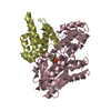
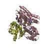
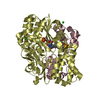
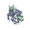
- PDB-2ik8: Crystal structure of the heterodimeric complex of human RGS16 and... -
+ Open data

- Basic information
| Entry | Database: PDB / ID: 2ik8 | ||||||
|---|---|---|---|---|---|---|---|
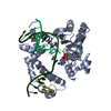
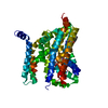
| Title | Crystal structure of the heterodimeric complex of human RGS16 and activated Gi alpha 1 | ||||||
 Components Components |
| ||||||
 Keywords Keywords |  SIGNALING PROTEIN / G protein signalling / RGS / heterotrimeric G protein / signalling complex / Structural Genomics / Structural Genomics Consortium / SGC SIGNALING PROTEIN / G protein signalling / RGS / heterotrimeric G protein / signalling complex / Structural Genomics / Structural Genomics Consortium / SGC | ||||||
| Function / homology |  Function and homology information Function and homology informationregulation of G protein-coupled receptor signaling pathway / Adenylate cyclase inhibitory pathway / positive regulation of protein localization to cell cortex / regulation of cAMP-mediated signaling / D2 dopamine receptor binding / G protein-coupled serotonin receptor binding / negative regulation of signal transduction / regulation of mitotic spindle organization / cellular response to forskolin / adenylate cyclase-inhibiting G protein-coupled receptor signaling pathway ...regulation of G protein-coupled receptor signaling pathway / Adenylate cyclase inhibitory pathway / positive regulation of protein localization to cell cortex / regulation of cAMP-mediated signaling / D2 dopamine receptor binding / G protein-coupled serotonin receptor binding / negative regulation of signal transduction / regulation of mitotic spindle organization / cellular response to forskolin / adenylate cyclase-inhibiting G protein-coupled receptor signaling pathway / visual perception / GTPase activator activity / Regulation of insulin secretion / G protein-coupled receptor binding / G-protein beta/gamma-subunit complex binding / adenylate cyclase-modulating G protein-coupled receptor signaling pathway / ADP signalling through P2Y purinoceptor 12 / response to peptide hormone / Adrenaline,noradrenaline inhibits insulin secretion / positive regulation of GTPase activity / G alpha (z) signalling events / ADORA2B mediated anti-inflammatory cytokines production / GPER1 signaling / GDP binding / heterotrimeric G-protein complex / cell cortex / midbody / G alpha (i) signalling events / G alpha (s) signalling events / G alpha (q) signalling events / Extra-nuclear estrogen signaling / calmodulin binding / cell cycle / G protein-coupled receptor signaling pathway / lysosomal membrane / cell division / GTPase activity / centrosome / nucleolus / GTP binding / magnesium ion binding / extracellular exosome / nucleoplasm / membrane / plasma membrane / cytoplasmSimilarity search - Function | ||||||
| Biological species |  Homo sapiens (human) Homo sapiens (human) | ||||||
| Method | X-RAY DIFFRACTION / SYNCHROTRON / MOLECULAR REPLACEMENT / Resolution: 2.71 Å | ||||||
 Authors Authors | Soundararajan, M. / Turnbull, A.P. / Papagrigoriou, E. / Debreczeni, J. / Gorrec, F. / von Delft, F. / Weigelt, J. / Edwards, A. / Arrowsmith, C. / Sundstrom, M. ...Soundararajan, M. / Turnbull, A.P. / Papagrigoriou, E. / Debreczeni, J. / Gorrec, F. / von Delft, F. / Weigelt, J. / Edwards, A. / Arrowsmith, C. / Sundstrom, M. / Doyle, D.A. / Structural Genomics Consortium (SGC) | ||||||
 Citation Citation | Journal: Proc.Natl.Acad.Sci.Usa / Year: 2008 Title: Structural diversity in the RGS domain and its interaction with heterotrimeric G protein alpha-subunits. Authors: Soundararajan, M. / Willard, F.S. / Kimple, A.J. / Turnbull, A.P. / Ball, L.J. / Schoch, G.A. / Gileadi, C. / Fedorov, O.Y. / Dowler, E.F. / Higman, V.A. / Hutsell, S.Q. / Sundstrom, M. / ...Authors: Soundararajan, M. / Willard, F.S. / Kimple, A.J. / Turnbull, A.P. / Ball, L.J. / Schoch, G.A. / Gileadi, C. / Fedorov, O.Y. / Dowler, E.F. / Higman, V.A. / Hutsell, S.Q. / Sundstrom, M. / Doyle, D.A. / Siderovski, D.P. | ||||||
| History |
|
- Structure visualization
| Structure viewer | Molecule: MolmilJmol/JSmol |
|---|
- Downloads & links
-Download
| PDBx/mmCIF format | 2ik8.cif.gz | 183.1 KB | Display | PDBx/mmCIF format |
|---|---|---|---|---|
| PDB format | pdb2ik8.ent.gz | 141.5 KB | Display | PDB format |
| PDBx/mmJSON format | 2ik8.json.gz | Tree view | PDBx/mmJSON format | |
| Others |  Other downloads Other downloads |
-Validation report
| Arichive directory | https://data.pdbj.org/pub/pdb/validation_reports/ik/2ik8ftp://data.pdbj.org/pub/pdb/validation_reports/ik/2ik8 | HTTPS FTP |
|---|
-Related structure data
| Related structure data | 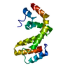 1zv4C 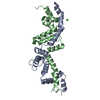 2a72C  2af0C 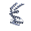 2bt2C 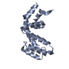 2bv1C 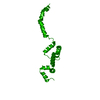 2es0C 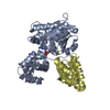 2gtpSC 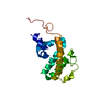 2i59C 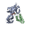 2ihbC  2ihdC  2jm5C  2jnuC 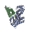 2odeC 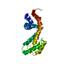 2owiC S: Starting model for refinement C: citing same article ( |
|---|---|
| Similar structure data |
-Links
 PDBj
PDBj


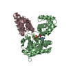
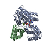
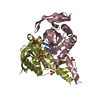
- Assembly
| Deposited unit | 
| |||||||||||||||||||||||||||||||||||||||||||||||||||||||||||||||||||||||||||||||||||||||||||||||||||||
|---|---|---|---|---|---|---|---|---|---|---|---|---|---|---|---|---|---|---|---|---|---|---|---|---|---|---|---|---|---|---|---|---|---|---|---|---|---|---|---|---|---|---|---|---|---|---|---|---|---|---|---|---|---|---|---|---|---|---|---|---|---|---|---|---|---|---|---|---|---|---|---|---|---|---|---|---|---|---|---|---|---|---|---|---|---|---|---|---|---|---|---|---|---|---|---|---|---|---|---|---|---|---|
| 1 | 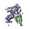
| |||||||||||||||||||||||||||||||||||||||||||||||||||||||||||||||||||||||||||||||||||||||||||||||||||||
| 2 | 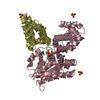
| |||||||||||||||||||||||||||||||||||||||||||||||||||||||||||||||||||||||||||||||||||||||||||||||||||||
| Unit cell |
| |||||||||||||||||||||||||||||||||||||||||||||||||||||||||||||||||||||||||||||||||||||||||||||||||||||
| Noncrystallographic symmetry (NCS) | NCS domain:
NCS domain segments: Refine code: 5
NCS ensembles :
|
-Components
-Protein , 2 types, 4 molecules ACBD
| #1: Protein | Mass: 37102.297 Da / Num. of mol.: 2 Source method: isolated from a genetically manipulated source Source: (gene. exp.) Homo sapiens (human) / Gene: GNAI1 / Plasmid: pNIC28-Bsa4 / Production host:  Escherichia coli (E. coli) / Strain (production host): BL21(DE3)R3 / References: UniProt: P63096 Escherichia coli (E. coli) / Strain (production host): BL21(DE3)R3 / References: UniProt: P63096#2: Protein | Mass: 15917.886 Da / Num. of mol.: 2 Source method: isolated from a genetically manipulated source Source: (gene. exp.) Homo sapiens (human) / Gene: RGS16, RGSR / Plasmid: pNIC28-Bsa4 / Production host: Escherichia coli (E. coli) / Strain (production host): BL21(DE3)R3 / References: UniProt: O15492 |
|---|
-Non-polymers , 5 types, 50 molecules 


| #3: Chemical |  Mass: 102.975 Da / Num. of mol.: 2 / Source method: obtained synthetically / Formula: AlF4 Mass: 102.975 Da / Num. of mol.: 2 / Source method: obtained synthetically / Formula: AlF4#4: Chemical |  Mass: 24.305 Da / Num. of mol.: 2 / Source method: obtained synthetically / Formula: Mg Mass: 24.305 Da / Num. of mol.: 2 / Source method: obtained synthetically / Formula: Mg#5: Chemical | ChemComp-SO4 / Sulfate Mass: 96.063 Da / Num. of mol.: 8 / Source method: obtained synthetically / Formula: SO4 Mass: 96.063 Da / Num. of mol.: 8 / Source method: obtained synthetically / Formula: SO4#6: Chemical | Guanosine diphosphate Type: RNA linking / Mass: 443.201 Da / Num. of mol.: 2 / Source method: obtained synthetically / Formula: C10H15N5O11P2 / Comment: GDP, energy-carrying molecule*YM Type: RNA linking / Mass: 443.201 Da / Num. of mol.: 2 / Source method: obtained synthetically / Formula: C10H15N5O11P2 / Comment: GDP, energy-carrying molecule*YM#7: Water | ChemComp-HOH / | WaterMass: 18.015 Da / Num. of mol.: 36 / Source method: isolated from a natural source / Formula: H2O |
|---|
-Experimental details
-Experiment
| Experiment | Method: X-RAY DIFFRACTION / Number of used crystals: 1 |
|---|
- Sample preparation
| Crystal | Density Matthews: 2.52 Å3/Da / Density % sol: 51.21 % |
|---|---|
| Crystal grow | Temperature: 293 K / Method: vapor diffusion, sitting drop / pH: 5.5 Details: 0.2M (NH4)2SO4, 0.1M Bis-Tris, pH 5.5, 25% PEG3350 , VAPOR DIFFUSION, SITTING DROP, temperature 293K |
-Data collection
| Diffraction | Mean temperature: 100 K |
|---|---|
| Diffraction source | Source: SYNCHROTRON / Site: SLS  / Beamline: X10SA / Wavelength: 0.9198 Å / Beamline: X10SA / Wavelength: 0.9198 Å |
| Detector | Type: MARMOSAIC 225 mm CCD / Detector: CCD / Date: Mar 24, 2006 |
| Radiation | Monochromator: Si (111) / Protocol: SINGLE WAVELENGTH / Monochromatic (M) / Laue (L): M / Scattering type: x-ray |
| Radiation wavelength | Wavelength: 0.9198 Å / Relative weight: 1 |
| Reflection | Resolution: 2.7→50 Å / Num. all: 29438 / Num. obs: 27900 / % possible obs: 99.3 % / Observed criterion σ(F): 0 / Observed criterion σ(I): 0 |
| Reflection shell | Resolution: 2.7→2.8 Å / % possible all: 99.2 |
- Processing
| Software |
| ||||||||||||||||||||||||||||||||||||||||||||||||||||||||||||||||||||||||||||||||||||||||||||||||||||||||||||||||||||||||||||||||||||||||||||||||||||||||||||||||||||||||||
|---|---|---|---|---|---|---|---|---|---|---|---|---|---|---|---|---|---|---|---|---|---|---|---|---|---|---|---|---|---|---|---|---|---|---|---|---|---|---|---|---|---|---|---|---|---|---|---|---|---|---|---|---|---|---|---|---|---|---|---|---|---|---|---|---|---|---|---|---|---|---|---|---|---|---|---|---|---|---|---|---|---|---|---|---|---|---|---|---|---|---|---|---|---|---|---|---|---|---|---|---|---|---|---|---|---|---|---|---|---|---|---|---|---|---|---|---|---|---|---|---|---|---|---|---|---|---|---|---|---|---|---|---|---|---|---|---|---|---|---|---|---|---|---|---|---|---|---|---|---|---|---|---|---|---|---|---|---|---|---|---|---|---|---|---|---|---|---|---|---|---|---|
| Refinement | Method to determine structure: MOLECULAR REPLACEMENT Starting model: PDB ID = 2GTP Resolution: 2.71→49.6 Å / Cor.coef. Fo:Fc: 0.928 / Cor.coef. Fo:Fc free: 0.895 / SU B: 31.847 / SU ML: 0.317 / TLS residual ADP flag: LIKELY RESIDUAL / Cross valid method: THROUGHOUT / σ(F): 0 / σ(I): 0 / ESU R: 2.095 / ESU R Free: 0.398 / Stereochemistry target values: MAXIMUM LIKELIHOOD / Details: HYDROGENS HAVE BEEN ADDED IN THE RIDING POSITIONS
| ||||||||||||||||||||||||||||||||||||||||||||||||||||||||||||||||||||||||||||||||||||||||||||||||||||||||||||||||||||||||||||||||||||||||||||||||||||||||||||||||||||||||||
| Solvent computation | Ion probe radii: 0.8 Å / Shrinkage radii: 0.8 Å / VDW probe radii: 1.4 Å / Solvent model: MASK | ||||||||||||||||||||||||||||||||||||||||||||||||||||||||||||||||||||||||||||||||||||||||||||||||||||||||||||||||||||||||||||||||||||||||||||||||||||||||||||||||||||||||||
| Displacement parameters | Biso mean: 52.189 Å2
| ||||||||||||||||||||||||||||||||||||||||||||||||||||||||||||||||||||||||||||||||||||||||||||||||||||||||||||||||||||||||||||||||||||||||||||||||||||||||||||||||||||||||||
| Refinement step | Cycle: LAST / Resolution: 2.71→49.6 Å
| ||||||||||||||||||||||||||||||||||||||||||||||||||||||||||||||||||||||||||||||||||||||||||||||||||||||||||||||||||||||||||||||||||||||||||||||||||||||||||||||||||||||||||
| Refine LS restraints |
| ||||||||||||||||||||||||||||||||||||||||||||||||||||||||||||||||||||||||||||||||||||||||||||||||||||||||||||||||||||||||||||||||||||||||||||||||||||||||||||||||||||||||||
| Refine LS restraints NCS | Dom-ID: 1 / Refine-ID: X-RAY DIFFRACTION
| ||||||||||||||||||||||||||||||||||||||||||||||||||||||||||||||||||||||||||||||||||||||||||||||||||||||||||||||||||||||||||||||||||||||||||||||||||||||||||||||||||||||||||
| LS refinement shell | Resolution: 2.71→2.781 Å / Total num. of bins used: 20
| ||||||||||||||||||||||||||||||||||||||||||||||||||||||||||||||||||||||||||||||||||||||||||||||||||||||||||||||||||||||||||||||||||||||||||||||||||||||||||||||||||||||||||
| Refinement TLS params. | Method: refined / Refine-ID: X-RAY DIFFRACTION
| ||||||||||||||||||||||||||||||||||||||||||||||||||||||||||||||||||||||||||||||||||||||||||||||||||||||||||||||||||||||||||||||||||||||||||||||||||||||||||||||||||||||||||
| Refinement TLS group |
|
