 Movie
Movie Controller


+ Open data

- Basic information
| Entry | Database: PDB / ID: 2i88 | ||||||
|---|---|---|---|---|---|---|---|
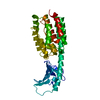
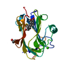
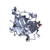
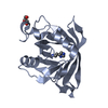
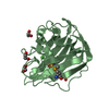
| Title | Crystal structure of the Channel-forming Domain of Colicin E1 | ||||||
 Components Components | Colicin-E1 | ||||||
 Keywords Keywords |  MEMBRANE PROTEIN / protein-membrane interactions / toxin-membrane interactions / toxin structure / voltage-gated channel / colicin MEMBRANE PROTEIN / protein-membrane interactions / toxin-membrane interactions / toxin structure / voltage-gated channel / colicin | ||||||
| Function / homology |  Function and homology information Function and homology informationnegative regulation of ion transmembrane transporter activity / pore-forming activity / killing of cells of another organism / defense response to Gram-negative bacterium / transmembrane transporter binding / plasma membraneSimilarity search - Function | ||||||
| Biological species |  Escherichia coli (E. coli) Escherichia coli (E. coli) | ||||||
| Method | X-RAY DIFFRACTION / MIR / Resolution: 2.5 Å | ||||||
 Authors Authors | Elkins, P. / Bunker, A. / Cramer, W.A. / Stauffacher, C.V. | ||||||
 Citation Citation | Journal: Structure / Year: 1997 Title: A mechanism for toxin insertion into membranes is suggested by the crystal structure of the channel-forming domain of colicin E1 Authors: Elkins, P. / Bunker, A. / Cramer, W.A. / Stauffacher, C.V. | ||||||
| History |
|
- Structure visualization
| Structure viewer | Molecule: MolmilJmol/JSmol |
|---|
- Downloads & links
-Download
| PDBx/mmCIF format | 2i88.cif.gz | 45.8 KB | Display | PDBx/mmCIF format |
|---|---|---|---|---|
| PDB format | pdb2i88.ent.gz | 32.8 KB | Display | PDB format |
| PDBx/mmJSON format | 2i88.json.gz | Tree view | PDBx/mmJSON format | |
| Others |  Other downloads Other downloads |
-Validation report
| Arichive directory | https://data.pdbj.org/pub/pdb/validation_reports/i8/2i88ftp://data.pdbj.org/pub/pdb/validation_reports/i8/2i88 | HTTPS FTP |
|---|
-Related structure data
| Related structure data | |
|---|---|
| Similar structure data |


-Links
 PDBj
PDBj- Assembly
| Deposited unit | 
| ||||||||
|---|---|---|---|---|---|---|---|---|---|
| 1 |
| ||||||||
| Unit cell |
|
-Components
| #1: Protein | Mass: 21196.420 Da / Num. of mol.: 1 / Fragment: C-terminal domain Source method: isolated from a genetically manipulated source Source: (gene. exp.) Escherichia coli (E. coli) / Strain: DM1187 / Gene: cea / Plasmid: pSKHY / Production host: Escherichia coli (E. coli) / References: UniProt: P02978 |
|---|---|
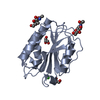
| #2: Water | ChemComp-HOH / Water Mass: 18.015 Da / Num. of mol.: 22 / Source method: isolated from a natural source / Formula: H2O Mass: 18.015 Da / Num. of mol.: 22 / Source method: isolated from a natural source / Formula: H2O |
-Experimental details
-Experiment
| Experiment | Method: X-RAY DIFFRACTION / Number of used crystals: 1 |
|---|
- Sample preparation
| Crystal | Density Matthews: 2.67 Å3/Da / Density % sol: 53.86 % |
|---|---|
| Crystal grow | Temperature: 295 K / Method: vapor diffusion Details: 50 mM MES at pH 6.0 25 mM Tris-HCl pH 7-8 100 mM NaCo or NaNO3 23-27% PEG 3350 very sensitive to temperatue stabilized by slow addition to of PEG 20,000 to 2-3%, VAPOR DIFFUSION, temperature 295K |
-Data collection
| Diffraction | Mean temperature: 295 K |
|---|---|
| Diffraction source | Source: ROTATING ANODE / Type: RIGAKU RU200 / Wavelength: 1.5418 |
| Detector | Type: RIGAKU RAXIS II / Detector: IMAGE PLATE |
| Radiation | Protocol: SINGLE WAVELENGTH / Monochromatic (M) / Laue (L): M / Scattering type: x-ray |
| Radiation wavelength | Wavelength: 1.5418 Å / Relative weight: 1 |
| Reflection | Resolution: 2.5→32 Å / Num. all: 7727 / Num. obs: 6684 / % possible obs: 86.5 % / Observed criterion σ(F): 1 / Observed criterion σ(I): 3 / Redundancy: 3.7 % / Biso Wilson estimate: 51.5 Å2 / Rsym value: 0.035 / Net I/σ(I): 13.97 |
| Reflection shell | Resolution: 2.5→2.64 Å / Redundancy: 2.2 % / Rmerge(I) obs: 0.125 / Mean I/σ(I) obs: 5.5 / Num. unique all: 683 / % possible all: 61.4 |
- Processing
| Software |
| ||||||||||||||||||||||||||||
|---|---|---|---|---|---|---|---|---|---|---|---|---|---|---|---|---|---|---|---|---|---|---|---|---|---|---|---|---|---|
| Refinement | Method to determine structure: MIR / Resolution: 2.5→32 Å / σ(F): 1 / σ(I): 3 / Stereochemistry target values: Engh & Huber
| ||||||||||||||||||||||||||||
| Displacement parameters | Biso mean: 38.885 Å2 | ||||||||||||||||||||||||||||
| Refinement step | Cycle: LAST / Resolution: 2.5→32 Å
| ||||||||||||||||||||||||||||
| Refine LS restraints |
|
