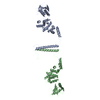
 Movie
Movie Controller


+ Open data

- Basic information
| Entry | Database: PDB / ID: 1cii | ||||||
|---|---|---|---|---|---|---|---|
| Title | COLICIN IA | ||||||
 Components Components | COLICIN IA | ||||||
 Keywords Keywords |  TRANSMEMBRANE PROTEIN / COLICIN / BACTERIOCIN / ION CHANNEL FORMATION TRANSMEMBRANE PROTEIN / COLICIN / BACTERIOCIN / ION CHANNEL FORMATION | ||||||
| Function / homology |  Function and homology information Function and homology informationpore-forming activity / killing of cells of another organism / defense response to Gram-negative bacterium / plasma membraneSimilarity search - Function | ||||||
| Biological species |  Escherichia coli (E. coli) Escherichia coli (E. coli) | ||||||
| Method | X-RAY DIFFRACTION / SYNCHROTRON / MIR / Resolution: 3 Å | ||||||
 Authors Authors | Wiener, M. / Freymann, D. / Ghosh, P. / Stroud, R. | ||||||
 Citation Citation | Journal: Nature / Year: 1997 Title: Crystal structure of colicin Ia. Authors: Wiener, M. / Freymann, D. / Ghosh, P. / Stroud, R.M. #1: Journal: Nat.Struct.Biol. / Year: 1994Title: The Domain Structure of the Ion Channel-Forming Protein Colicin Ia Authors: Ghosh, P. / Mel, S.F. / Stroud, R.M. | ||||||
| History |
|
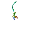
- Structure visualization
| Structure viewer | Molecule: MolmilJmol/JSmol |
|---|
- Downloads & links
-Download
| PDBx/mmCIF format | 1cii.cif.gz | 127.9 KB | Display | PDBx/mmCIF format |
|---|---|---|---|---|
| PDB format | pdb1cii.ent.gz | 100.1 KB | Display | PDB format |
| PDBx/mmJSON format | 1cii.json.gz | Tree view | PDBx/mmJSON format | |
| Others |  Other downloads Other downloads |
-Validation report
| Arichive directory | https://data.pdbj.org/pub/pdb/validation_reports/ci/1ciiftp://data.pdbj.org/pub/pdb/validation_reports/ci/1cii | HTTPS FTP |
|---|
-Related structure data
| Similar structure data |
|---|


-Links
 PDBj
PDBj- Assembly
| Deposited unit | 
| ||||||||
|---|---|---|---|---|---|---|---|---|---|
| 1 |
| ||||||||
| Unit cell |
|
-Components
| #1: Protein | Mass: 67036.273 Da / Num. of mol.: 1 Source method: isolated from a genetically manipulated source Source: (gene. exp.) Escherichia coli (E. coli) / Strain: 294 / Gene: CIA / Plasmid: PJK5 / Gene (production host): CIA / Production host: Escherichia coli (E. coli) / Strain (production host): 294 / References: UniProt: P06716 |
|---|---|
| Compound details | TRANSLOCATION (T) DOMAIN: RESIDUES 23 - 225. RECEPTOR-BINDING (R) DOMAIN: RESIDUES 282 - 385. ...TRANSLOCAT |
-Experimental details
-Experiment
| Experiment | Method: X-RAY DIFFRACTION / Number of used crystals: 3 |
|---|
- Sample preparation
| Crystal | Density Matthews: 5.9 Å3/Da / Density % sol: 78 % | ||||||||||||||||||||||||||||||||||||||||||||||||
|---|---|---|---|---|---|---|---|---|---|---|---|---|---|---|---|---|---|---|---|---|---|---|---|---|---|---|---|---|---|---|---|---|---|---|---|---|---|---|---|---|---|---|---|---|---|---|---|---|---|
| Crystal grow | Method: vapor diffusion - hanging drop - streak seeding / pH: 5.2 Details: PROTEIN WAS CRYSTALLIZED BY HANGING-DROP VAPOR DIFFUSION AGAINST RESERVOIRS OF 1.0 M NH4(SO4)2, 20 MM NA-CITRATE (PH 5.2), 200 MM NACL, STARTING WITH 6 MICROLITERS OF PROTEIN AT 2 MG/ML IN ...Details: PROTEIN WAS CRYSTALLIZED BY HANGING-DROP VAPOR DIFFUSION AGAINST RESERVOIRS OF 1.0 M NH4(SO4)2, 20 MM NA-CITRATE (PH 5.2), 200 MM NACL, STARTING WITH 6 MICROLITERS OF PROTEIN AT 2 MG/ML IN 20 MM NA-CITRATE (PH 5.2), 200 MM NACL, 5 MM DTT, AND 4 MICROLITERS RESERVOIR SOLUTION. THESE DROPS OF MUTANT PROTEIN WERE STREAK-SEEDED FROM STOCKS GENERATED FROM CRUSHED DIFFRACTION-QUALITY WILD-TYPE COLICIN IA CRYSTALS. CRYSTALS WERE HARVESTED TO 1.1 M NA2SO4, 200 MM NACL, 20 MM NA-CITRATE (PH 5.2), AND 5 MM DTT. FOR DERIVATIZATION, CRYSTALS WERE WASHED IN DTT-FREE HARVEST BUFFER AND SOAKED FOR ONE WEEK IN 1 MM CH3HGCL., vapor diffusion - hanging drop - streak seeding | ||||||||||||||||||||||||||||||||||||||||||||||||
| Crystal grow | *PLUS Method: vapor diffusion, hanging drop | ||||||||||||||||||||||||||||||||||||||||||||||||
| Components of the solutions | *PLUS
|
-Data collection
| Diffraction | Mean temperature: 90 K |
|---|---|
| Diffraction source | Source: SYNCHROTRON / Site: SSRL  / Beamline: BL7-1 / Wavelength: 1.08 / Beamline: BL7-1 / Wavelength: 1.08 |
| Detector | Type: MAR scanner 300 mm plate / Detector: IMAGE PLATE / Date: Apr 1, 1994 / Details: PT-COATED FUSED SILICA MIRROR |
| Radiation | Monochromator: SI(111) / Monochromatic (M) / Laue (L): M / Scattering type: x-ray |
| Radiation wavelength | Wavelength: 1.08 Å / Relative weight: 1 |
| Reflection | Resolution: 3→25 Å / Num. obs: 28266 / % possible obs: 84 % / Observed criterion σ(I): 0 / Redundancy: 3.4 % / Rmerge(I) obs: 0.13 / Rsym value: 0.13 / Net I/σ(I): 5.7 |
| Reflection shell | Resolution: 3→3.11 Å / Redundancy: 2.3 % / Rmerge(I) obs: 0.309 / Mean I/σ(I) obs: 1.7 / Rsym value: 0.309 / % possible all: 73 |
| Reflection shell | *PLUS % possible obs: 73 % |
- Processing
| Software |
| ||||||||||||||||||||||||||||||||||||||||||||||||||||||||||||||||||||||||||||||||
|---|---|---|---|---|---|---|---|---|---|---|---|---|---|---|---|---|---|---|---|---|---|---|---|---|---|---|---|---|---|---|---|---|---|---|---|---|---|---|---|---|---|---|---|---|---|---|---|---|---|---|---|---|---|---|---|---|---|---|---|---|---|---|---|---|---|---|---|---|---|---|---|---|---|---|---|---|---|---|---|---|---|
| Refinement | Method to determine structure: MIR / Resolution: 3→8 Å / Rfactor Rfree error: 0.007 / Data cutoff high absF: 100000 / Data cutoff low absF: 0.1 / Isotropic thermal model: INDIVIDUAL RESTRAINED / Cross valid method: THROUGHOUT / σ(F): 0 Details: ANISOTROPIC B-FACTOR CORRECTION APPLIED TO THE DIFFRACTION DATA. BULK SOLVENT MASK CALCULATED AND UTILIZED.
| ||||||||||||||||||||||||||||||||||||||||||||||||||||||||||||||||||||||||||||||||
| Displacement parameters | Biso mean: 66.5 Å2 | ||||||||||||||||||||||||||||||||||||||||||||||||||||||||||||||||||||||||||||||||
| Refine analyze | Luzzati d res low obs: 25 Å / Luzzati sigma a obs: 0.34 Å | ||||||||||||||||||||||||||||||||||||||||||||||||||||||||||||||||||||||||||||||||
| Refinement step | Cycle: LAST / Resolution: 3→8 Å
| ||||||||||||||||||||||||||||||||||||||||||||||||||||||||||||||||||||||||||||||||
| Refine LS restraints |
| ||||||||||||||||||||||||||||||||||||||||||||||||||||||||||||||||||||||||||||||||
| LS refinement shell | Resolution: 3→3.1 Å / Rfactor Rfree error: 0.03 / Total num. of bins used: 10
| ||||||||||||||||||||||||||||||||||||||||||||||||||||||||||||||||||||||||||||||||
| Xplor file |
| ||||||||||||||||||||||||||||||||||||||||||||||||||||||||||||||||||||||||||||||||
| Software | *PLUS Name: X-PLOR / Version: 3.1 / Classification: refinement | ||||||||||||||||||||||||||||||||||||||||||||||||||||||||||||||||||||||||||||||||
| Refine LS restraints | *PLUS
|
