 Movie
Movie Controller


+ Open data

- Basic information
| Entry | Database: PDB / ID: 2he0 | ||||||
|---|---|---|---|---|---|---|---|
| Title | Crystal structure of a human Notch1 ankyrin domain mutant | ||||||
 Components Components | Notch1 preproprotein variant | ||||||
 Keywords Keywords |  SIGNALING PROTEIN / Notch / Ankyrin / signalling SIGNALING PROTEIN / Notch / Ankyrin / signalling | ||||||
| Function / homology |  Function and homology information Function and homology informationDefective LFNG causes SCDO3 / coronary sinus valve morphogenesis / cardiac right atrium morphogenesis / cardiac right ventricle formation / growth involved in heart morphogenesis / Notch signaling pathway involved in regulation of secondary heart field cardioblast proliferation / cell differentiation in spinal cord / retinal cone cell differentiation / venous endothelial cell differentiation / arterial endothelial cell differentiation ...Defective LFNG causes SCDO3 / coronary sinus valve morphogenesis / cardiac right atrium morphogenesis / cardiac right ventricle formation / growth involved in heart morphogenesis / Notch signaling pathway involved in regulation of secondary heart field cardioblast proliferation / cell differentiation in spinal cord / retinal cone cell differentiation / venous endothelial cell differentiation / arterial endothelial cell differentiation / cardiac chamber formation / epithelial cell fate commitment / negative regulation of pro-B cell differentiation / Pre-NOTCH Processing in the Endoplasmic Reticulum / negative regulation of inner ear auditory receptor cell differentiation / mitral valve formation / cell migration involved in endocardial cushion formation / glomerular mesangial cell development / negative regulation of photoreceptor cell differentiation / negative regulation of cell proliferation involved in heart valve morphogenesis / regulation of somitogenesis / inhibition of neuroepithelial cell differentiation / endocardium morphogenesis / atrioventricular node development / foregut morphogenesis / regulation of cell adhesion involved in heart morphogenesis / distal tubule development / MAML1-RBP-Jkappa- ICN1 complex / regulation of epithelial cell proliferation involved in prostate gland development / auditory receptor cell fate commitment / positive regulation of aorta morphogenesis / negative regulation of endothelial cell chemotaxis / neuroendocrine cell differentiation / collecting duct development / negative regulation of extracellular matrix constituent secretion / positive regulation of transcription of Notch receptor target / cellular response to tumor cell / positive regulation of apoptotic process involved in morphogenesis / compartment pattern specification / vasculogenesis involved in coronary vascular morphogenesis / T-helper 17 type immune response / regulation of extracellular matrix assembly / endocardial cell differentiation / epithelial to mesenchymal transition involved in endocardial cushion formation / cardiac ventricle morphogenesis / Constitutive Signaling by NOTCH1 t(7;9)(NOTCH1:M1580_K2555) Translocation Mutant / positive regulation of smooth muscle cell differentiation / cardiac left ventricle morphogenesis / mesenchymal cell development / epidermal cell fate specification / coronary vein morphogenesis / negative regulation of collagen biosynthetic process / cardiac vascular smooth muscle cell development / negative regulation of myotube differentiation / somatic stem cell division / left/right axis specification / negative regulation of cardiac muscle hypertrophy / negative regulation of cell adhesion molecule production / interleukin-17-mediated signaling pathway / positive regulation of endothelial cell differentiation / secretory columnal luminar epithelial cell differentiation involved in prostate glandular acinus development / endocardium development / apoptotic process involved in embryonic digit morphogenesis / positive regulation of cardiac epithelial to mesenchymal transition / Pre-NOTCH Processing in Golgi / cardiac epithelial to mesenchymal transition / negative regulation of calcium ion-dependent exocytosis / cardiac muscle cell myoblast differentiation / cellular response to follicle-stimulating hormone stimulus / pericardium morphogenesis / cardiac atrium morphogenesis / negative regulation of catalytic activity / neuronal stem cell population maintenance / tissue regeneration / regulation of stem cell proliferation / negative regulation of oligodendrocyte differentiation / positive regulation of astrocyte differentiation / calcium-ion regulated exocytosis / pulmonary valve morphogenesis / heart trabecula morphogenesis / negative regulation of biomineral tissue development / endoderm development / coronary artery morphogenesis / negative regulation of cell-cell adhesion mediated by cadherin / prostate gland epithelium morphogenesis / luteolysis / cardiac muscle tissue morphogenesis / ventricular trabecula myocardium morphogenesis / negative regulation of myoblast differentiation / negative regulation of cell migration involved in sprouting angiogenesis / transcription regulator activator activity / positive regulation of BMP signaling pathway / tube formation / negative regulation of stem cell differentiation / Loss of Function of FBXW7 in Cancer and NOTCH1 Signaling / positive regulation of keratinocyte differentiation / astrocyte differentiation / inflammatory response to antigenic stimulus / positive regulation of Ras protein signal transduction / negative regulation of ossificationSimilarity search - Function | ||||||
| Biological species |  Homo sapiens (human) Homo sapiens (human) | ||||||
| Method | X-RAY DIFFRACTION / SYNCHROTRON / MOLECULAR REPLACEMENT / Resolution: 1.9 Å | ||||||
 Authors Authors | Gupta, D. / Ehebauer, M.T. / Chirgadze, D.Y. / Martinez Arias, A. / Blundell, T.L. | ||||||
 Citation Citation | Journal: TO BE PUBLISHED Title: Crystal structure of a human Notch1 ankyrin domain mutant Authors: Gupta, D. / Ehebauer, M.T. / Chirgadze, D.Y. / Martinez Arias, A. / Blundell, T.L. | ||||||
| History |
|
- Structure visualization
| Structure viewer | Molecule: MolmilJmol/JSmol |
|---|
- Downloads & links
-Download
| PDBx/mmCIF format | 2he0.cif.gz | 97.1 KB | Display | PDBx/mmCIF format |
|---|---|---|---|---|
| PDB format | pdb2he0.ent.gz | 73 KB | Display | PDB format |
| PDBx/mmJSON format | 2he0.json.gz | Tree view | PDBx/mmJSON format | |
| Others |  Other downloads Other downloads |
-Validation report
| Arichive directory | https://data.pdbj.org/pub/pdb/validation_reports/he/2he0ftp://data.pdbj.org/pub/pdb/validation_reports/he/2he0 | HTTPS FTP |
|---|
-Related structure data
| Related structure data |  1yyhS S: Starting model for refinement |
|---|---|
| Similar structure data |


-Links
 PDBj
PDBj


- Assembly
| Deposited unit | 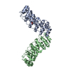
| ||||||||
|---|---|---|---|---|---|---|---|---|---|
| 1 |
| ||||||||
| Unit cell |
|
-Components
| #1: Protein | Mass: 27634.691 Da / Num. of mol.: 2 / Fragment: Ankyrin Domain, residues 1873-2115 / Mutation: E58A, R66A Source method: isolated from a genetically manipulated source Source: (gene. exp.) Homo sapiens (human) / Gene: Notch 1 / Plasmid: pET41a / Production host:  Escherichia coli (E. coli) / Strain (production host): Rosetta (DE3) / References: UniProt: P46531 Escherichia coli (E. coli) / Strain (production host): Rosetta (DE3) / References: UniProt: P46531#2: Chemical | ChemComp-EDO / | Ethylene glycol  Mass: 62.068 Da / Num. of mol.: 1 / Source method: obtained synthetically / Formula: C2H6O2 Mass: 62.068 Da / Num. of mol.: 1 / Source method: obtained synthetically / Formula: C2H6O2#3: Water | ChemComp-HOH / | Water Mass: 18.015 Da / Num. of mol.: 416 / Source method: isolated from a natural source / Formula: H2O Mass: 18.015 Da / Num. of mol.: 416 / Source method: isolated from a natural source / Formula: H2O |
|---|
-Experimental details
-Experiment
| Experiment | Method: X-RAY DIFFRACTION / Number of used crystals: 1 |
|---|
- Sample preparation
| Crystal | Density Matthews: 3.26 Å3/Da / Density % sol: 62.32 % |
|---|---|
| Crystal grow | Temperature: 298 K / Method: vapor diffusion, hanging drop / pH: 8 Details: 0.55M Sodium/Potassium Tartrate, pH 8.0, VAPOR DIFFUSION, HANGING DROP, temperature 298K |
-Data collection
| Diffraction | Mean temperature: 100 K |
|---|---|
| Diffraction source | Source: SYNCHROTRON / Site: ESRF  / Beamline: ID14-1 / Wavelength: 0.9769 Å / Beamline: ID14-1 / Wavelength: 0.9769 Å |
| Detector | Type: ADSC QUANTUM 315 / Detector: CCD / Date: May 19, 2006 |
| Radiation | Protocol: SINGLE WAVELENGTH / Monochromatic (M) / Laue (L): M / Scattering type: x-ray |
| Radiation wavelength | Wavelength: 0.9769 Å / Relative weight: 1 |
| Reflection | Resolution: 1.9→50 Å / Num. all: 45594 / Num. obs: 45547 / % possible obs: 99.9 % / Observed criterion σ(F): 0 / Observed criterion σ(I): 3 / Redundancy: 10.7 % / Biso Wilson estimate: 26.6 Å2 / Rmerge(I) obs: 0.066 / Rsym value: 0.066 / Net I/σ(I): 10.6 |
| Reflection shell | Resolution: 1.9→1.94 Å / Rmerge(I) obs: 0.488 / Rsym value: 0.488 / % possible all: 99.9 |
- Processing
| Software |
| ||||||||||||||||||||||||||||||||||||||||||||||||||||||||||||||||||||||||||||||||||||||||||
|---|---|---|---|---|---|---|---|---|---|---|---|---|---|---|---|---|---|---|---|---|---|---|---|---|---|---|---|---|---|---|---|---|---|---|---|---|---|---|---|---|---|---|---|---|---|---|---|---|---|---|---|---|---|---|---|---|---|---|---|---|---|---|---|---|---|---|---|---|---|---|---|---|---|---|---|---|---|---|---|---|---|---|---|---|---|---|---|---|---|---|---|
| Refinement | Method to determine structure: MOLECULAR REPLACEMENT Starting model: PDB ENTRY 1YYH Resolution: 1.9→48.51 Å / Cor.coef. Fo:Fc: 0.949 / Cor.coef. Fo:Fc free: 0.924 / SU B: 2.961 / SU ML: 0.089 / Cross valid method: THROUGHOUT / σ(F): 0 / ESU R: 0.124 / ESU R Free: 0.127 / Stereochemistry target values: MAXIMUM LIKELIHOOD / Details: HYDROGENS HAVE BEEN ADDED IN THE RIDING POSITIONS
| ||||||||||||||||||||||||||||||||||||||||||||||||||||||||||||||||||||||||||||||||||||||||||
| Solvent computation | Ion probe radii: 0.8 Å / Shrinkage radii: 0.8 Å / VDW probe radii: 1.2 Å / Solvent model: MASK | ||||||||||||||||||||||||||||||||||||||||||||||||||||||||||||||||||||||||||||||||||||||||||
| Displacement parameters | Biso mean: 31.288 Å2
| ||||||||||||||||||||||||||||||||||||||||||||||||||||||||||||||||||||||||||||||||||||||||||
| Refinement step | Cycle: LAST / Resolution: 1.9→48.51 Å
| ||||||||||||||||||||||||||||||||||||||||||||||||||||||||||||||||||||||||||||||||||||||||||
| Refine LS restraints |
| ||||||||||||||||||||||||||||||||||||||||||||||||||||||||||||||||||||||||||||||||||||||||||
| LS refinement shell | Resolution: 1.901→1.951 Å / Total num. of bins used: 20
|
