 Movie
Movie Controller


+ Open data

- Basic information
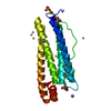
| Entry | Database: PDB / ID: 2qc9 | ||||||
|---|---|---|---|---|---|---|---|
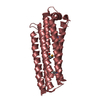
| Title | Mouse Notch 1 Ankyrin Repeat Intracellular Domain | ||||||
 Components Components | Notch 1 protein | ||||||
 Keywords Keywords |  TRANSCRIPTION / beta-hydroxy asparagine / ankyrin repeat / factor inhibiting HIF TRANSCRIPTION / beta-hydroxy asparagine / ankyrin repeat / factor inhibiting HIF | ||||||
| Function / homology |  Function and homology information Function and homology informationPre-NOTCH Processing in Golgi / regulation of cardioblast proliferation / regulation of inner ear auditory receptor cell differentiation / positive regulation of ephrin receptor signaling pathway / positive regulation of glial cell differentiation / osteoblast fate commitment / venous blood vessel morphogenesis / Activated NOTCH1 Transmits Signal to the Nucleus / coronary sinus valve morphogenesis / cardiac right atrium morphogenesis ...Pre-NOTCH Processing in Golgi / regulation of cardioblast proliferation / regulation of inner ear auditory receptor cell differentiation / positive regulation of ephrin receptor signaling pathway / positive regulation of glial cell differentiation / osteoblast fate commitment / venous blood vessel morphogenesis / Activated NOTCH1 Transmits Signal to the Nucleus / coronary sinus valve morphogenesis / cardiac right atrium morphogenesis / cardiac right ventricle formation / growth involved in heart morphogenesis / Notch signaling pathway involved in regulation of secondary heart field cardioblast proliferation / cell differentiation in spinal cord / retinal cone cell differentiation / venous endothelial cell differentiation / arterial endothelial cell differentiation / cardiac chamber formation / epithelial cell fate commitment / negative regulation of pro-B cell differentiation / negative regulation of inner ear auditory receptor cell differentiation / mitral valve formation / cell migration involved in endocardial cushion formation / glomerular mesangial cell development / negative regulation of photoreceptor cell differentiation / negative regulation of cell proliferation involved in heart valve morphogenesis / regulation of somitogenesis / inhibition of neuroepithelial cell differentiation / endocardium morphogenesis / atrioventricular node development / foregut morphogenesis / regulation of cell adhesion involved in heart morphogenesis / distal tubule development / MAML1-RBP-Jkappa- ICN1 complex / regulation of epithelial cell proliferation involved in prostate gland development / auditory receptor cell fate commitment / positive regulation of aorta morphogenesis / negative regulation of endothelial cell chemotaxis / NOTCH1 Intracellular Domain Regulates Transcription / RUNX3 regulates NOTCH signaling / neuroendocrine cell differentiation / collecting duct development / negative regulation of extracellular matrix constituent secretion / positive regulation of transcription of Notch receptor target / cellular response to tumor cell / positive regulation of apoptotic process involved in morphogenesis / Notch-HLH transcription pathway / compartment pattern specification / vasculogenesis involved in coronary vascular morphogenesis / T-helper 17 type immune response / endocardial cushion development / regulation of extracellular matrix assembly / endocardial cell differentiation / epithelial to mesenchymal transition involved in endocardial cushion formation / cardiac ventricle morphogenesis / positive regulation of smooth muscle cell differentiation / cardiac left ventricle morphogenesis / mesenchymal cell development / epidermal cell fate specification / regulation of Notch signaling pathway / coronary vein morphogenesis / negative regulation of collagen biosynthetic process / cardiac vascular smooth muscle cell development / negative regulation of myotube differentiation / somatic stem cell division / left/right axis specification / negative regulation of cardiac muscle hypertrophy / negative regulation of cell adhesion molecule production / interleukin-17-mediated signaling pathway / positive regulation of endothelial cell differentiation / secretory columnal luminar epithelial cell differentiation involved in prostate glandular acinus development / endocardium development / apoptotic process involved in embryonic digit morphogenesis / glial cell differentiation / positive regulation of cardiac epithelial to mesenchymal transition / cardiac epithelial to mesenchymal transition / negative regulation of calcium ion-dependent exocytosis / cardiac muscle cell myoblast differentiation / cellular response to follicle-stimulating hormone stimulus / neuron fate commitment / pericardium morphogenesis / cardiac atrium morphogenesis / negative regulation of catalytic activity / neuronal stem cell population maintenance / tissue regeneration / regulation of stem cell proliferation / negative regulation of oligodendrocyte differentiation / positive regulation of astrocyte differentiation / calcium-ion regulated exocytosis / pulmonary valve morphogenesis / heart trabecula morphogenesis / negative regulation of biomineral tissue development / endoderm development / coronary artery morphogenesis / negative regulation of cell-cell adhesion mediated by cadherin / prostate gland epithelium morphogenesis / luteolysis / cardiac muscle tissue morphogenesis / ventricular trabecula myocardium morphogenesis / negative regulation of myoblast differentiationSimilarity search - Function | ||||||
| Biological species |  Mus musculus (house mouse) Mus musculus (house mouse) | ||||||
| Method | X-RAY DIFFRACTION / MOLECULAR REPLACEMENT / Resolution: 2.35 Å | ||||||
 Authors Authors | McDonough, M.A. / Schofield, C.J. | ||||||
 Citation Citation | Journal: J.Biol.Chem. / Year: 2007 Title: Asparaginyl hydroxylation of the Notch ankyrin repeat domain by factor inhibiting hypoxia-inducible factor. Authors: Coleman, M.L. / McDonough, M.A. / Hewitson, K.S. / Coles, C. / Mecinovic, J. / Edelmann, M. / Cook, K.M. / Cockman, M.E. / Lancaster, D.E. / Kessler, B.M. / Oldham, N.J. / Ratcliffe, P.J. / Schofield, C.J. | ||||||
| History |
| ||||||
| Remark 300 | BIOMOLECULE: 1 THIS ENTRY CONTAINS THE CRYSTALLOGRAPHIC ASYMMETRIC UNIT WHICH CONSISTS OF 2 CHAIN(S) ...BIOMOLECULE: 1 THIS ENTRY CONTAINS THE CRYSTALLOGRAPHIC ASYMMETRIC UNIT WHICH CONSISTS OF 2 CHAIN(S). THE AUTHOR REPORTS THE BIOLOGICAL ASSEMBLY TO BE UNKNOWN |
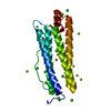
- Structure visualization
| Structure viewer | Molecule: MolmilJmol/JSmol |
|---|
- Downloads & links
-Download
| PDBx/mmCIF format | 2qc9.cif.gz | 97.5 KB | Display | PDBx/mmCIF format |
|---|---|---|---|---|
| PDB format | pdb2qc9.ent.gz | 73.2 KB | Display | PDB format |
| PDBx/mmJSON format | 2qc9.json.gz | Tree view | PDBx/mmJSON format | |
| Others |  Other downloads Other downloads |
-Validation report
| Arichive directory | https://data.pdbj.org/pub/pdb/validation_reports/qc/2qc9ftp://data.pdbj.org/pub/pdb/validation_reports/qc/2qc9 | HTTPS FTP |
|---|
-Related structure data
| Related structure data |  3p3nC  3p3pC  1yyhS S: Starting model for refinement C: citing same article ( |
|---|---|
| Similar structure data |


-Links
 PDBj
PDBj


- Assembly
| Deposited unit | 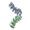
| ||||||||
|---|---|---|---|---|---|---|---|---|---|
| 1 | 
| ||||||||
| 2 | 
| ||||||||
| Unit cell |
| ||||||||
| Details | THE AUTHOR REPORTS THE BIOLOGICAL ASSEMBLY TO BE UNKNOWN |
-Components
| #1: Protein | Mass: 23132.811 Da / Num. of mol.: 2 / Fragment: Ankyrin Repeat Domain Source method: isolated from a genetically manipulated source Source: (gene. exp.) Mus musculus (house mouse) / Gene: Notch1 / Plasmid: pGEX-4T1 / Species (production host): Escherichia coli / Production host:  Escherichia coli BL21(DE3) (bacteria) / Strain (production host): BL21 (DE3) / References: UniProt: Q8K428, UniProt: Q01705*PLUS Escherichia coli BL21(DE3) (bacteria) / Strain (production host): BL21 (DE3) / References: UniProt: Q8K428, UniProt: Q01705*PLUS#2: Water | ChemComp-HOH / | Water Mass: 18.015 Da / Num. of mol.: 442 / Source method: isolated from a natural source / Formula: H2O Mass: 18.015 Da / Num. of mol.: 442 / Source method: isolated from a natural source / Formula: H2O |
|---|
-Experimental details
-Experiment
| Experiment | Method: X-RAY DIFFRACTION / Number of used crystals: 1 |
|---|
- Sample preparation
| Crystal | Density Matthews: 3.47 Å3/Da / Density % sol: 64.52 % |
|---|---|
| Crystal grow | Temperature: 298 K / Method: vapor diffusion, sitting drop / pH: 7 Details: 30% PEG 6000, 100mM HEPES, pH 7.0, VAPOR DIFFUSION, SITTING DROP, temperature 298.0K |
-Data collection
| Diffraction | Mean temperature: 105 K |
|---|---|
| Diffraction source | Source: ROTATING ANODE / Type: BRUKER AXS MICROSTAR / Wavelength: 1.5418 Å |
| Detector | Type: BRUKER SMART 6000 / Detector: CCD / Date: Dec 15, 2005 / Details: Montel 200 |
| Radiation | Protocol: SINGLE WAVELENGTH / Monochromatic (M) / Laue (L): M / Scattering type: x-ray |
| Radiation wavelength | Wavelength: 1.5418 Å / Relative weight: 1 |
| Reflection twin | Type: hemihedral / Operator: h,-h-k,-l / Fraction: 0.19 |
| Reflection | Resolution: 2.35→67 Å / Num. obs: 48746 / % possible obs: 100 % / Redundancy: 11.1 % / Rsym value: 0.1423 / Net I/σ(I): 7.43 |
| Reflection shell | Resolution: 2.35→2.4 Å / Redundancy: 9.27 % / Mean I/σ(I) obs: 2.56 / Num. unique all: 1534 / Rsym value: 0.4344 / % possible all: 100 |
- Processing
| Software |
| ||||||||||||||||||||||||||||||||||||||||||||||||||||||
|---|---|---|---|---|---|---|---|---|---|---|---|---|---|---|---|---|---|---|---|---|---|---|---|---|---|---|---|---|---|---|---|---|---|---|---|---|---|---|---|---|---|---|---|---|---|---|---|---|---|---|---|---|---|---|---|
| Refinement | Method to determine structure: MOLECULAR REPLACEMENT Starting model: 1YYH Resolution: 2.35→67 Å / σ(F): 2205
| ||||||||||||||||||||||||||||||||||||||||||||||||||||||
| Solvent computation | Bsol: 63.82 Å2 | ||||||||||||||||||||||||||||||||||||||||||||||||||||||
| Displacement parameters | Biso mean: 21.372 Å2
| ||||||||||||||||||||||||||||||||||||||||||||||||||||||
| Refinement step | Cycle: LAST / Resolution: 2.35→67 Å
| ||||||||||||||||||||||||||||||||||||||||||||||||||||||
| Refine LS restraints |
| ||||||||||||||||||||||||||||||||||||||||||||||||||||||
| LS refinement shell | Refine-ID: X-RAY DIFFRACTION / Total num. of bins used: 8
| ||||||||||||||||||||||||||||||||||||||||||||||||||||||
| Xplor file |
|
