 Movie
Movie Controller


+ Open data

- Basic information
| Entry | Database: PDB / ID: 2h61 | ||||||
|---|---|---|---|---|---|---|---|
| Title | X-ray structure of human Ca2+-loaded S100B | ||||||
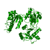
 Components Components | (Protein S100-B) x 2 | ||||||
 Keywords Keywords | METAL BINDING PROTEIN /  SIGNALING PROTEIN / S100 / EF-hand / Calcium-binding / RAGE SIGNALING PROTEIN / S100 / EF-hand / Calcium-binding / RAGE | ||||||
| Function / homology |  Function and homology informationadaptive thermogenesis / sympathetic neuron projection extension / RAGE receptor binding / ion binding / S100 protein binding / regulation of neuronal synaptic plasticity / TRAF6 mediated NF-kB activation / Advanced glycosylation endproduct receptor signaling / Nuclear signaling by ERBB4 / ruffle ...adaptive thermogenesis / sympathetic neuron projection extension / RAGE receptor binding / ion binding / S100 protein binding / regulation of neuronal synaptic plasticity / TRAF6 mediated NF-kB activation / Advanced glycosylation endproduct receptor signaling / Nuclear signaling by ERBB4 / ruffle / positive regulation of neuron differentiation / axonogenesis / central nervous system development / tau protein binding / TAK1-dependent IKK and NF-kappa-B activation / memory / calcium-dependent protein binding / positive regulation of canonical NF-kappaB signal transduction / learning or memory / cell adhesion / intracellular membrane-bounded organelle / neuronal cell body / calcium ion binding / positive regulation of cell population proliferation / perinuclear region of cytoplasm / protein homodimerization activity / extracellular space / zinc ion binding / extracellular region / nucleoplasm / identical protein binding / nucleus / cytosol / cytoplasm Function and homology informationadaptive thermogenesis / sympathetic neuron projection extension / RAGE receptor binding / ion binding / S100 protein binding / regulation of neuronal synaptic plasticity / TRAF6 mediated NF-kB activation / Advanced glycosylation endproduct receptor signaling / Nuclear signaling by ERBB4 / ruffle ...adaptive thermogenesis / sympathetic neuron projection extension / RAGE receptor binding / ion binding / S100 protein binding / regulation of neuronal synaptic plasticity / TRAF6 mediated NF-kB activation / Advanced glycosylation endproduct receptor signaling / Nuclear signaling by ERBB4 / ruffle / positive regulation of neuron differentiation / axonogenesis / central nervous system development / tau protein binding / TAK1-dependent IKK and NF-kappa-B activation / memory / calcium-dependent protein binding / positive regulation of canonical NF-kappaB signal transduction / learning or memory / cell adhesion / intracellular membrane-bounded organelle / neuronal cell body / calcium ion binding / positive regulation of cell population proliferation / perinuclear region of cytoplasm / protein homodimerization activity / extracellular space / zinc ion binding / extracellular region / nucleoplasm / identical protein binding / nucleus / cytosol / cytoplasmSimilarity search - Function | ||||||
| Biological species |  Homo sapiens (human) Homo sapiens (human) | ||||||
| Method | X-RAY DIFFRACTION / MOLECULAR REPLACEMENT / Resolution: 1.9 Å | ||||||
 Authors Authors | Ostendorp, T. / Heizmann, C.W. / Kroneck, P.M.H. / Fritz, G. | ||||||
 Citation Citation | Journal: Embo J. / Year: 2007 Title: Structural and functional insights into RAGE activation by multimeric S100B. Authors: Ostendorp, T. / Leclerc, E. / Galichet, A. / Koch, M. / Demling, N. / Weigle, B. / Heizmann, C.W. / Kroneck, P.M. / Fritz, G. | ||||||
| History |
| ||||||
| Remark 999 | SEQUENCE The crystals of S100B were dissolved and checked by reversed phase HPLC and mass ...SEQUENCE The crystals of S100B were dissolved and checked by reversed phase HPLC and mass spectrometry. It was found that about 30% of the N-terminal Met in S100B was post translationally modified by E.coli. The mass deviation is in agreement with FME. |
- Structure visualization
| Structure viewer | Molecule: MolmilJmol/JSmol |
|---|
- Downloads & links
-Download
| PDBx/mmCIF format | 2h61.cif.gz | 177.2 KB | Display | PDBx/mmCIF format |
|---|---|---|---|---|
| PDB format | pdb2h61.ent.gz | 140.7 KB | Display | PDB format |
| PDBx/mmJSON format | 2h61.json.gz | Tree view | PDBx/mmJSON format | |
| Others |  Other downloads Other downloads |
-Validation report
| Arichive directory | https://data.pdbj.org/pub/pdb/validation_reports/h6/2h61ftp://data.pdbj.org/pub/pdb/validation_reports/h6/2h61 | HTTPS FTP |
|---|
-Related structure data
| Related structure data |  1mhoS S: Starting model for refinement |
|---|---|
| Similar structure data |


-Links
 PDBj
PDBj


- Assembly
| Deposited unit | 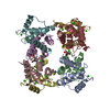
| ||||||||
|---|---|---|---|---|---|---|---|---|---|
| 1 |
| ||||||||
| Unit cell |
| ||||||||
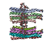
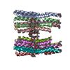
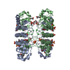
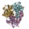
| Details | The assymetric unit contains four homodimers which associate to two tetramers, which combine to an octamer |
-Components
| #1: Protein | Mass: 10727.037 Da / Num. of mol.: 5 Source method: isolated from a genetically manipulated source Details: MET at the N-terminus / Source: (gene. exp.) Homo sapiens (human) / Plasmid: pGEMEX / Species (production host): Escherichia coli / Production host:  Escherichia coli BL21(DE3) (bacteria) / Strain (production host): BL21(DE3) / References: UniProt: P04271 Escherichia coli BL21(DE3) (bacteria) / Strain (production host): BL21(DE3) / References: UniProt: P04271#2: Protein | Mass: 10755.047 Da / Num. of mol.: 3 Source method: isolated from a genetically manipulated source Details: FME at the N-terminus / Source: (gene. exp.) Homo sapiens (human) / Plasmid: pGEMEX / Species (production host): Escherichia coli / Production host: Escherichia coli BL21(DE3) (bacteria) / Strain (production host): BL21(DE3) / References: UniProt: P04271#3: Chemical | ChemComp-CA /   Mass: 40.078 Da / Num. of mol.: 18 / Source method: obtained synthetically / Formula: Ca Mass: 40.078 Da / Num. of mol.: 18 / Source method: obtained synthetically / Formula: Ca#4: Chemical | ChemComp-PG4 / | Polyethylene glycol  Mass: 194.226 Da / Num. of mol.: 1 / Source method: obtained synthetically / Formula: C8H18O5 / Comment: precipitant*YM Mass: 194.226 Da / Num. of mol.: 1 / Source method: obtained synthetically / Formula: C8H18O5 / Comment: precipitant*YM#5: Water | ChemComp-HOH / | Water Mass: 18.015 Da / Num. of mol.: 617 / Source method: isolated from a natural source / Formula: H2O Mass: 18.015 Da / Num. of mol.: 617 / Source method: isolated from a natural source / Formula: H2O |
|---|
-Experimental details
-Experiment
| Experiment | Method: X-RAY DIFFRACTION / Number of used crystals: 1 |
|---|
- Sample preparation
| Crystal | Density Matthews: 2.06 Å3/Da / Density % sol: 40.26 % |
|---|---|
| Crystal grow | Temperature: 289 K / Method: vapor diffusion, sitting drop / pH: 7.2 Details: 0.1 M HEPES, 18-20%PEG400, 0.2 M CaCl2, pH 7.2, VAPOR DIFFUSION, SITTING DROP, temperature 289K |
-Data collection
| Diffraction | Mean temperature: 100 K |
|---|---|
| Diffraction source | Source: ROTATING ANODE / Type: SIEMENS / Wavelength: 1.54 |
| Detector | Type: MAR scanner 345 mm plate / Detector: IMAGE PLATE |
| Radiation | Protocol: SINGLE WAVELENGTH / Monochromatic (M) / Laue (L): M / Scattering type: x-ray |
| Radiation wavelength | Wavelength: 1.54 Å / Relative weight: 1 |
| Reflection | Resolution: 1.9→50 Å / Num. obs: 63871 / % possible obs: 99 % / Observed criterion σ(F): 2 / Observed criterion σ(I): 2 / Rmerge(I) obs: 0.078 / Net I/σ(I): 15.8 |
| Reflection shell | Resolution: 1.9→2 Å / Rmerge(I) obs: 0.315 / Mean I/σ(I) obs: 4.9 / Num. unique all: 7334 / % possible all: 94.1 |
- Processing
| Software |
| |||||||||||||||||||||||||||||||||||||||||||||||||||||||||||||||||||||||||||||||||||||||||||||||||||||||||||||||||||||||||||||||||||||||||||||||||||||||||||||||||||||||||||||||||||||||||||||||||||||||||||||||||||||||||||||||||
|---|---|---|---|---|---|---|---|---|---|---|---|---|---|---|---|---|---|---|---|---|---|---|---|---|---|---|---|---|---|---|---|---|---|---|---|---|---|---|---|---|---|---|---|---|---|---|---|---|---|---|---|---|---|---|---|---|---|---|---|---|---|---|---|---|---|---|---|---|---|---|---|---|---|---|---|---|---|---|---|---|---|---|---|---|---|---|---|---|---|---|---|---|---|---|---|---|---|---|---|---|---|---|---|---|---|---|---|---|---|---|---|---|---|---|---|---|---|---|---|---|---|---|---|---|---|---|---|---|---|---|---|---|---|---|---|---|---|---|---|---|---|---|---|---|---|---|---|---|---|---|---|---|---|---|---|---|---|---|---|---|---|---|---|---|---|---|---|---|---|---|---|---|---|---|---|---|---|---|---|---|---|---|---|---|---|---|---|---|---|---|---|---|---|---|---|---|---|---|---|---|---|---|---|---|---|---|---|---|---|---|---|---|---|---|---|---|---|---|---|---|---|---|---|---|---|---|
| Refinement | Method to determine structure: MOLECULAR REPLACEMENT Starting model: pdb entry 1MHO Resolution: 1.9→30.32 Å / Cor.coef. Fo:Fc: 0.96 / Cor.coef. Fo:Fc free: 0.921 / SU B: 7.638 / SU ML: 0.122 / TLS residual ADP flag: LIKELY RESIDUAL / Cross valid method: THROUGHOUT / σ(F): 3.54 / ESU R: 0.169 / ESU R Free: 0.165 / Stereochemistry target values: MAXIMUM LIKELIHOOD / Details: HYDROGENS HAVE BEEN ADDED IN THE RIDING POSITIONS
| |||||||||||||||||||||||||||||||||||||||||||||||||||||||||||||||||||||||||||||||||||||||||||||||||||||||||||||||||||||||||||||||||||||||||||||||||||||||||||||||||||||||||||||||||||||||||||||||||||||||||||||||||||||||||||||||||
| Solvent computation | Ion probe radii: 0.8 Å / Shrinkage radii: 0.8 Å / VDW probe radii: 1.2 Å / Solvent model: MASK | |||||||||||||||||||||||||||||||||||||||||||||||||||||||||||||||||||||||||||||||||||||||||||||||||||||||||||||||||||||||||||||||||||||||||||||||||||||||||||||||||||||||||||||||||||||||||||||||||||||||||||||||||||||||||||||||||
| Displacement parameters | Biso mean: 27.082 Å2
| |||||||||||||||||||||||||||||||||||||||||||||||||||||||||||||||||||||||||||||||||||||||||||||||||||||||||||||||||||||||||||||||||||||||||||||||||||||||||||||||||||||||||||||||||||||||||||||||||||||||||||||||||||||||||||||||||
| Refinement step | Cycle: LAST / Resolution: 1.9→30.32 Å
| |||||||||||||||||||||||||||||||||||||||||||||||||||||||||||||||||||||||||||||||||||||||||||||||||||||||||||||||||||||||||||||||||||||||||||||||||||||||||||||||||||||||||||||||||||||||||||||||||||||||||||||||||||||||||||||||||
| Refine LS restraints |
| |||||||||||||||||||||||||||||||||||||||||||||||||||||||||||||||||||||||||||||||||||||||||||||||||||||||||||||||||||||||||||||||||||||||||||||||||||||||||||||||||||||||||||||||||||||||||||||||||||||||||||||||||||||||||||||||||
| LS refinement shell | Resolution: 1.9→1.949 Å / Total num. of bins used: 20
| |||||||||||||||||||||||||||||||||||||||||||||||||||||||||||||||||||||||||||||||||||||||||||||||||||||||||||||||||||||||||||||||||||||||||||||||||||||||||||||||||||||||||||||||||||||||||||||||||||||||||||||||||||||||||||||||||
| Refinement TLS params. | Method: refined / Refine-ID: X-RAY DIFFRACTION
| |||||||||||||||||||||||||||||||||||||||||||||||||||||||||||||||||||||||||||||||||||||||||||||||||||||||||||||||||||||||||||||||||||||||||||||||||||||||||||||||||||||||||||||||||||||||||||||||||||||||||||||||||||||||||||||||||
| Refinement TLS group | Refine-ID: X-RAY DIFFRACTION / Selection: ALL / Auth seq-ID: 0 - 89 / Label seq-ID: 1 - 90
|
