UDP-3-O-acyl-N-acetylglucosamine deacetylase / UDP-3-O-[3-hydroxymyristoyl] N-acetylglucosamine deacetylase activity / UDP-3-O-acyl-N-acetylglucosamine deacetylase activity / lipid A biosynthetic process / metal ion binding Similarity search - Function
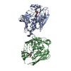
BIOMOLECULE: 1, 2 THIS ENTRY CONTAINS THE CRYSTALLOGRAPHIC ASYMMETRIC UNIT WHICH CONSISTS OF 2 ...BIOMOLECULE: 1, 2 THIS ENTRY CONTAINS THE CRYSTALLOGRAPHIC ASYMMETRIC UNIT WHICH CONSISTS OF 2 CHAIN(S). ALTHOUGH IN THE CRYSTAL THIS MOLECULE APPEARS TO FORM A DIMER, THE MONOMER IS BIOLOGICALLY ACTIVE. SEE REMARK 350 FOR INFORMATION ON GENERATING THE BIOLOGICAL MOLECULE(S).
Remark 999
SEQUENCE THE RESIDUE NUMBERING SCHEME FOR THIS STRUCTURE FOLLOWS THAT OF THE E. COLI ENZYME. THIS ...SEQUENCE THE RESIDUE NUMBERING SCHEME FOR THIS STRUCTURE FOLLOWS THAT OF THE E. COLI ENZYME. THIS TREATMENT RESULTS IN A BREAK IN THE SEQUENTIAL NUMBERING IN A COUPLE OF PLACES.
Mass: 18.015 Da / Num. of mol.: 56 / Source method: isolated from a natural source / Formula: H2O
-
Experimental details
-
Experiment
Experiment
Method: X-RAY DIFFRACTION / Number of used crystals: 1
-
Sample preparation
Crystal
Density Matthews: 2.91 Å3/Da / Density % sol: 57.77 %
Crystal grow
Temperature: 294 K / Method: vapor diffusion, hanging drop / pH: 7.5 Details: 100 mM HEPES (pH 7.5), 180 mM NaCl, 14% PEG 3350, 5 mM ZnSO4, VAPOR DIFFUSION, HANGING DROP, temperature 294K
-
Data collection
Diffraction
Mean temperature: 200 K
Diffraction source
Source: SYNCHROTRON / Site: CHESS / Beamline: F1 / Wavelength: 0.9124 Å
Detector
Type: ADSC QUANTUM 4 / Detector: CCD / Date: Feb 10, 2006 / Details: Rh coated Si monochromatic mirror
Radiation
Monochromator: Horizontal bent Si(111), asymmetrically cut with water cooled Cu Block Protocol: SINGLE WAVELENGTH / Monochromatic (M) / Laue (L): M / Scattering type: x-ray
Radiation wavelength
Wavelength: 0.9124 Å / Relative weight: 1
Reflection
Resolution: 2.7→50 Å / Num. all: 19244 / Num. obs: 17601 / % possible obs: 97.7 % / Rmerge(I) obs: 0.069 / Net I/σ(I): 12.6
Reflection shell
Resolution: 2.7→2.8 Å / Mean I/σ(I) obs: 2.2 / % possible all: 97.2
In the structure databanks used in Yorodumi, some data are registered as the other names, "COVID-19 virus" and "2019-nCoV". Here are the details of the virus and the list of structure data.
Jan 31, 2019. EMDB accession codes are about to change! (news from PDBe EMDB page)
EMDB accession codes are about to change! (news from PDBe EMDB page)
The allocation of 4 digits for EMDB accession codes will soon come to an end. Whilst these codes will remain in use, new EMDB accession codes will include an additional digit and will expand incrementally as the available range of codes is exhausted. The current 4-digit format prefixed with “EMD-” (i.e. EMD-XXXX) will advance to a 5-digit format (i.e. EMD-XXXXX), and so on. It is currently estimated that the 4-digit codes will be depleted around Spring 2019, at which point the 5-digit format will come into force.
The EM Navigator/Yorodumi systems omit the EMD- prefix.
Related info.:Q: What is EMD? / ID/Accession-code notation in Yorodumi/EM Navigator
Yorodumi is a browser for structure data from EMDB, PDB, SASBDB, etc.
This page is also the successor to EM Navigator detail page, and also detail information page/front-end page for Omokage search.
The word "yorodu" (or yorozu) is an old Japanese word meaning "ten thousand". "mi" (miru) is to see.
Related info.:EMDB / PDB / SASBDB / Comparison of 3 databanks / Yorodumi Search / Aug 31, 2016. New EM Navigator & Yorodumi / Yorodumi Papers / Jmol/JSmol / Function and homology information / Changes in new EM Navigator and Yorodumi
 Movie
Movie Controller


 Open data
 Basic information
 Components
 Keywords
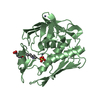
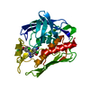
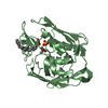
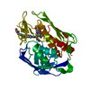
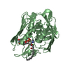
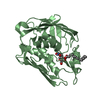
 HYDROLASE / LpxC-inhibitor complex
 Function and homology information

 Authors
 Citation
 Structure visualization
 Downloads & links
 Other downloads
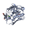


 PDBj
PDBj Assembly
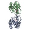


 Mass: 65.409 Da / Num. of mol.: 5 / Source method: obtained synthetically / Formula: Zn
 Mass: 35.453 Da / Num. of mol.: 1 / Source method: obtained synthetically / Formula: Cl
 Mass: 431.563 Da / Num. of mol.: 2 / Source method: obtained synthetically / Formula: C22H41NO7
 Mass: 18.015 Da / Num. of mol.: 56 / Source method: isolated from a natural source / Formula: H2O
 Sample preparation
 / Beamline: F1 / Wavelength: 0.9124 Å
 Processing