 Movie
Movie Controller


+ Open data

- Basic information
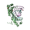
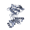
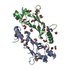
| Entry | Database: PDB / ID: 2fu5 | ||||||
|---|---|---|---|---|---|---|---|
| Title | structure of Rab8 in complex with MSS4 | ||||||
 Components Components |
| ||||||
 Keywords Keywords |  SIGNALING PROTEIN / MSS4:Rab8 protein complex / GEF:GTPase nucleotide free complex / nucleotide exchange via unfolding of nucleotide binding region SIGNALING PROTEIN / MSS4:Rab8 protein complex / GEF:GTPase nucleotide free complex / nucleotide exchange via unfolding of nucleotide binding region | ||||||
| Function / homology |  Function and homology information Function and homology informationVxPx cargo-targeting to cilium / TBC/RABGAPs / neurotransmitter receptor transport to postsynaptic membrane / RAB geranylgeranylation / RAB GEFs exchange GTP for GDP on RABs / small GTPase binding => GO:0031267 / Golgi vesicle fusion to target membrane / vesicle-mediated transport in synapse / regulation of protein transport / post-Golgi vesicle-mediated transport ...VxPx cargo-targeting to cilium / TBC/RABGAPs / neurotransmitter receptor transport to postsynaptic membrane / RAB geranylgeranylation / RAB GEFs exchange GTP for GDP on RABs / small GTPase binding => GO:0031267 / Golgi vesicle fusion to target membrane / vesicle-mediated transport in synapse / regulation of protein transport / post-Golgi vesicle-mediated transport / neurotransmitter receptor transport, endosome to postsynaptic membrane / Anchoring of the basal body to the plasma membrane / Regulation of PLK1 Activity at G2/M Transition / myosin V binding / vesicle docking involved in exocytosis / regulation of exocytosis / trans-Golgi network transport vesicle / non-motile cilium / ciliary base / small GTPase-mediated signal transduction / cilium assembly / protein secretion / phagocytic vesicle / centriole / axonogenesis / guanyl-nucleotide exchange factor activity / ciliary basal body / regulation of autophagy / protein localization to plasma membrane / regulation of long-term neuronal synaptic plasticity / cytoplasmic vesicle membrane / cilium / recycling endosome / autophagy / cellular response to insulin stimulus / recycling endosome membrane / phagocytic vesicle membrane / GDP binding / regulation of protein localization / synaptic vesicle / protein transport / midbody / cytoplasmic vesicle / dendritic spine / membrane fusion / postsynaptic density / endosome / GTPase activity / centrosome / neuronal cell body / glutamatergic synapse / synapse / dendrite / GTP binding / protein kinase binding / Golgi apparatus / zinc ion binding / membrane / plasma membrane / cytosolSimilarity search - Function | ||||||
| Biological species |  Homo sapiens (human) Homo sapiens (human) Mus musculus (house mouse) Mus musculus (house mouse) | ||||||
| Method | X-RAY DIFFRACTION / SYNCHROTRON / MOLECULAR REPLACEMENT / Resolution: 2 Å | ||||||
 Authors Authors | Itzen, A. / Pylypenko, O. / Goody, R.S. / Rak, A. | ||||||
 Citation Citation | Journal: Embo J. / Year: 2006 Title: Nucleotide exchange via local protein unfolding-structure of Rab8 in complex with MSS4 Authors: Itzen, A. / Pylypenko, O. / Goody, R.S. / Alexandrov, K. / Rak, A. | ||||||
| History |
|
- Structure visualization
| Structure viewer | Molecule: MolmilJmol/JSmol |
|---|
- Downloads & links
-Download
| PDBx/mmCIF format | 2fu5.cif.gz | 124 KB | Display | PDBx/mmCIF format |
|---|---|---|---|---|
| PDB format | pdb2fu5.ent.gz | 95.1 KB | Display | PDB format |
| PDBx/mmJSON format | 2fu5.json.gz | Tree view | PDBx/mmJSON format | |
| Others |  Other downloads Other downloads |
-Validation report
| Arichive directory | https://data.pdbj.org/pub/pdb/validation_reports/fu/2fu5ftp://data.pdbj.org/pub/pdb/validation_reports/fu/2fu5 | HTTPS FTP |
|---|
-Related structure data
| Related structure data | 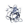 1hxrS S: Starting model for refinement |
|---|---|
| Similar structure data |


-Links
 PDBj
PDBj


- Assembly
| Deposited unit | 
| ||||||||
|---|---|---|---|---|---|---|---|---|---|
| 1 | 
| ||||||||
| 2 | 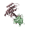
| ||||||||
| Unit cell |
|
-Components
| #1: Protein | Mass: 13198.093 Da / Num. of mol.: 2 / Fragment: residues 11-123 Source method: isolated from a genetically manipulated source Source: (gene. exp.) Homo sapiens (human) / Gene: RABIF, MSS4, RASGRF3 / Plasmid: pET19 / Production host:  Escherichia coli (E. coli) / Strain (production host): BL21(DE3)RIL / References: UniProt: P47224 Escherichia coli (E. coli) / Strain (production host): BL21(DE3)RIL / References: UniProt: P47224#2: Protein | Mass: 20953.076 Da / Num. of mol.: 2 / Fragment: residues 1-183 Source method: isolated from a genetically manipulated source Source: (gene. exp.) Mus musculus (house mouse) / Gene: Rab8a, Mel, Rab8 / Plasmid: pET30a / Production host: Escherichia coli (E. coli) / Strain (production host): BL21(DE3)RIL / References: UniProt: P55258#3: Chemical |   Mass: 65.409 Da / Num. of mol.: 2 / Source method: obtained synthetically / Formula: Zn Mass: 65.409 Da / Num. of mol.: 2 / Source method: obtained synthetically / Formula: Zn#4: Chemical | 2-Mercaptoethanol  Mass: 78.133 Da / Num. of mol.: 2 / Source method: obtained synthetically / Formula: C2H6OS Mass: 78.133 Da / Num. of mol.: 2 / Source method: obtained synthetically / Formula: C2H6OS#5: Water | ChemComp-HOH / | Water Mass: 18.015 Da / Num. of mol.: 301 / Source method: isolated from a natural source / Formula: H2O Mass: 18.015 Da / Num. of mol.: 301 / Source method: isolated from a natural source / Formula: H2O |
|---|
-Experimental details
-Experiment
| Experiment | Method: X-RAY DIFFRACTION / Number of used crystals: 1 |
|---|
- Sample preparation
| Crystal | Density Matthews: 2.41 Å3/Da / Density % sol: 48.96 % |
|---|---|
| Crystal grow | Temperature: 293 K / Method: vapor diffusion, hanging drop / pH: 9 Details: 20% PEG3350, 50mM TrisHCL pH 9.0, 0.2M magnesium acetate, 0.15% (w/v) 1,2,3-heptanetriole, VAPOR DIFFUSION, HANGING DROP, temperature 293K |
-Data collection
| Diffraction | Mean temperature: 100 K |
|---|---|
| Diffraction source | Source: SYNCHROTRON / Site: ESRF  / Beamline: ID14-2 / Wavelength: 0.934 Å / Beamline: ID14-2 / Wavelength: 0.934 Å |
| Detector | Type: ADSC QUANTUM 4 / Detector: CCD |
| Radiation | Protocol: SINGLE WAVELENGTH / Monochromatic (M) / Laue (L): M / Scattering type: x-ray |
| Radiation wavelength | Wavelength: 0.934 Å / Relative weight: 1 |
| Reflection | Resolution: 2→19.17 Å / Num. all: 40718 / Num. obs: 40718 / % possible obs: 94.5 % / Observed criterion σ(F): -3 / Observed criterion σ(I): -3 |
| Reflection shell | Resolution: 2→2.05 Å / % possible all: 74.6 |
- Processing
| Software |
| ||||||||||||||||||||||||||||||||||||||||||||||||||||||||||||||||||||||||||||||||||||||||||
|---|---|---|---|---|---|---|---|---|---|---|---|---|---|---|---|---|---|---|---|---|---|---|---|---|---|---|---|---|---|---|---|---|---|---|---|---|---|---|---|---|---|---|---|---|---|---|---|---|---|---|---|---|---|---|---|---|---|---|---|---|---|---|---|---|---|---|---|---|---|---|---|---|---|---|---|---|---|---|---|---|---|---|---|---|---|---|---|---|---|---|---|
| Refinement | Method to determine structure: MOLECULAR REPLACEMENT Starting model: PDB ENTRY 1hxr Resolution: 2→19.17 Å / Cor.coef. Fo:Fc: 0.945 / Cor.coef. Fo:Fc free: 0.903 / SU B: 3.901 / SU ML: 0.113 / Cross valid method: THROUGHOUT / σ(F): 0 / ESU R: 0.186 / ESU R Free: 0.175 / Stereochemistry target values: Engh & Huber
| ||||||||||||||||||||||||||||||||||||||||||||||||||||||||||||||||||||||||||||||||||||||||||
| Solvent computation | Ion probe radii: 0.8 Å / Shrinkage radii: 0.8 Å / VDW probe radii: 1.2 Å / Solvent model: BABINET MODEL WITH MASK | ||||||||||||||||||||||||||||||||||||||||||||||||||||||||||||||||||||||||||||||||||||||||||
| Displacement parameters | Biso mean: 44.426 Å2 | ||||||||||||||||||||||||||||||||||||||||||||||||||||||||||||||||||||||||||||||||||||||||||
| Refinement step | Cycle: LAST / Resolution: 2→19.17 Å
| ||||||||||||||||||||||||||||||||||||||||||||||||||||||||||||||||||||||||||||||||||||||||||
| Refine LS restraints |
| ||||||||||||||||||||||||||||||||||||||||||||||||||||||||||||||||||||||||||||||||||||||||||
| LS refinement shell | Resolution: 2→2.051 Å / Total num. of bins used: 20 /
|
