 Movie
Movie Controller

[English] 日本語
 Yorodumi
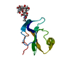
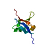
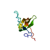
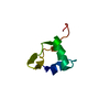
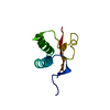
- PDB-2fk4: Solution structure of the C-terminal zinc binding domain of the H... -
+ Open data

- Basic information
| Entry | Database: PDB / ID: 2fk4 | ||||||
|---|---|---|---|---|---|---|---|
| Title | Solution structure of the C-terminal zinc binding domain of the HPV16 E6 oncoprotein | ||||||
 Components Components | Protein E6 | ||||||
 Keywords Keywords | Metal binding protein /  Oncoprotein / ZINC BINDING DOMAIN Oncoprotein / ZINC BINDING DOMAIN | ||||||
| Function / homology |  Function and homology information Function and homology informationsymbiont-mediated suppression of host transcription / regulation of proteolysis / activation of GTPase activity / PDZ domain binding / symbiont-mediated suppression of host cytoplasmic pattern recognition receptor signaling pathway via inhibition of IRF3 activity / symbiont-mediated perturbation of host ubiquitin-like protein modification / host cell cytoplasm / symbiont-mediated suppression of host type I interferon-mediated signaling pathway / virus-mediated perturbation of host defense response / DNA-templated transcription ...symbiont-mediated suppression of host transcription / regulation of proteolysis / activation of GTPase activity / PDZ domain binding / symbiont-mediated suppression of host cytoplasmic pattern recognition receptor signaling pathway via inhibition of IRF3 activity / symbiont-mediated perturbation of host ubiquitin-like protein modification / host cell cytoplasm / symbiont-mediated suppression of host type I interferon-mediated signaling pathway / virus-mediated perturbation of host defense response / DNA-templated transcription / host cell nucleus / positive regulation of transcription by RNA polymerase II / DNA binding / identical protein binding / metal ion bindingSimilarity search - Function | ||||||
| Biological species |  Human papillomavirus type 16 Human papillomavirus type 16 | ||||||
| Method | SOLUTION NMR / simulated annealing, protocol refine.inp | ||||||
 Authors Authors | Nomine, Y. / Charbonnier, S. | ||||||
 Citation Citation | Journal: Mol.Cell / Year: 2006 Title: Structural and functional analysis of E6 oncoprotein: insights in the molecular pathways of human papillomavirus-mediated pathogenesis Authors: Nomine, Y. / Masson, M. / Charbonnier, S. / Zanier, K. / Ristriani, T. / Deryckere, F. / Sibler, A.P. / Desplancq, D. / Atkinson, R.A. / Weiss, E. / Orfanoudakis, G. / Kieffer, B. / Trave, G. #1: Journal: J.Biomol.Nmr / Year: 2005 Title: 1H and 15N resonance assignment, secondary structure and dynamic behaviour of the C-terminal domain of human papillomavirus oncoprotein E6. Authors: Charbonnier, S. / Miguet, L. / Potier, N. / Van Dorsselaer, A. / Atkinson, R.A. / Kieffer, B. | ||||||
| History |
|
- Structure visualization
| Structure viewer | Molecule: MolmilJmol/JSmol |
|---|
- Downloads & links
-Download
| PDBx/mmCIF format | 2fk4.cif.gz | 250.8 KB | Display | PDBx/mmCIF format |
|---|---|---|---|---|
| PDB format | pdb2fk4.ent.gz | 207.8 KB | Display | PDB format |
| PDBx/mmJSON format | 2fk4.json.gz | Tree view | PDBx/mmJSON format | |
| Others |  Other downloads Other downloads |
-Validation report
| Arichive directory | https://data.pdbj.org/pub/pdb/validation_reports/fk/2fk4ftp://data.pdbj.org/pub/pdb/validation_reports/fk/2fk4 | HTTPS FTP |
|---|
-Related structure data
| Similar structure data |
|---|


-Links
 PDBj
PDBj
- Assembly
| Deposited unit | 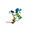
| |||||||||
|---|---|---|---|---|---|---|---|---|---|---|
| 1 |
| |||||||||
| NMR ensembles |
|
-Components
| #1: Protein | Mass: 8905.230 Da / Num. of mol.: 1 / Fragment: C-TERMINAL DOMAIN / Mutation: C4S, C20S, C34S, C63S Source method: isolated from a genetically manipulated source Source: (gene. exp.) Human papillomavirus type 16 / Genus: Alphapapillomavirus / Species: Human papillomavirus - 16 / Production host:  Escherichia coli (E. coli) / Strain (production host): BL21 DE2 / References: UniProt: P03126 Escherichia coli (E. coli) / Strain (production host): BL21 DE2 / References: UniProt: P03126 |
|---|---|
| #2: Chemical | ChemComp-ZN /   Mass: 65.409 Da / Num. of mol.: 1 / Source method: obtained synthetically / Formula: Zn Mass: 65.409 Da / Num. of mol.: 1 / Source method: obtained synthetically / Formula: Zn |
-Experimental details
-Experiment
| Experiment | Method: SOLUTION NMR | ||||||||||||
|---|---|---|---|---|---|---|---|---|---|---|---|---|---|
| NMR experiment |
| ||||||||||||
| NMR details | Text: This structure was determined using standard 2D homonuclear and 3D 15N heteronuclear techniques. |
- Sample preparation
| Details |
| |||||||||||||||
|---|---|---|---|---|---|---|---|---|---|---|---|---|---|---|---|---|
| Sample conditions |
|
-NMR measurement
| Radiation | Protocol: SINGLE WAVELENGTH / Monochromatic (M) / Laue (L): M | |||||||||||||||
|---|---|---|---|---|---|---|---|---|---|---|---|---|---|---|---|---|
| Radiation wavelength | Relative weight: 1 | |||||||||||||||
| NMR spectrometer |
|
- Processing
| NMR software |
| ||||||||||||||||||||
|---|---|---|---|---|---|---|---|---|---|---|---|---|---|---|---|---|---|---|---|---|---|
| Refinement | Method: simulated annealing, protocol refine.inp / Software ordinal: 1 Details: NOE distance restraints were split in two sets. One set involved residues that were identified as beeing affected by conformational exchange by relaxation dispersion experiments. These ...Details: NOE distance restraints were split in two sets. One set involved residues that were identified as beeing affected by conformational exchange by relaxation dispersion experiments. These distances were assigned an additional 1A to the upper distance limit. Reported close contacts are due to conformational heterogeneities in the set of NOES. The dynamical properties of this protein have been described in depth in the J. Biomol. NMR reference cited above. | ||||||||||||||||||||
| NMR representative | Selection criteria: fewest violations | ||||||||||||||||||||
| NMR ensemble | Conformer selection criteria: structures with the lowest energy Conformers calculated total number: 20 / Conformers submitted total number: 10 |
