 Movie
Movie Controller

[English] 日本語
 Yorodumi
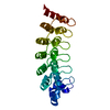
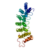
- PDB-2f8y: Crystal structure of human Notch1 ankyrin repeats to 1.55A resolution. -
+ Open data

- Basic information
| Entry | Database: PDB / ID: 2f8y | ||||||
|---|---|---|---|---|---|---|---|
| Title | Crystal structure of human Notch1 ankyrin repeats to 1.55A resolution. | ||||||
 Components Components | Notch homolog 1, translocation-associated (Drosophila) | ||||||
 Keywords Keywords |  TRANSCRIPTION / Notch / Ankyrin repeats TRANSCRIPTION / Notch / Ankyrin repeats | ||||||
| Function / homology |  Function and homology information Function and homology informationDefective LFNG causes SCDO3 / coronary sinus valve morphogenesis / cardiac right atrium morphogenesis / cardiac right ventricle formation / growth involved in heart morphogenesis / Notch signaling pathway involved in regulation of secondary heart field cardioblast proliferation / cell differentiation in spinal cord / retinal cone cell differentiation / venous endothelial cell differentiation / arterial endothelial cell differentiation ...Defective LFNG causes SCDO3 / coronary sinus valve morphogenesis / cardiac right atrium morphogenesis / cardiac right ventricle formation / growth involved in heart morphogenesis / Notch signaling pathway involved in regulation of secondary heart field cardioblast proliferation / cell differentiation in spinal cord / retinal cone cell differentiation / venous endothelial cell differentiation / arterial endothelial cell differentiation / cardiac chamber formation / epithelial cell fate commitment / negative regulation of pro-B cell differentiation / Pre-NOTCH Processing in the Endoplasmic Reticulum / negative regulation of inner ear auditory receptor cell differentiation / mitral valve formation / cell migration involved in endocardial cushion formation / glomerular mesangial cell development / negative regulation of photoreceptor cell differentiation / negative regulation of cell proliferation involved in heart valve morphogenesis / regulation of somitogenesis / inhibition of neuroepithelial cell differentiation / endocardium morphogenesis / atrioventricular node development / foregut morphogenesis / regulation of cell adhesion involved in heart morphogenesis / distal tubule development / MAML1-RBP-Jkappa- ICN1 complex / regulation of epithelial cell proliferation involved in prostate gland development / auditory receptor cell fate commitment / positive regulation of aorta morphogenesis / negative regulation of endothelial cell chemotaxis / neuroendocrine cell differentiation / collecting duct development / negative regulation of extracellular matrix constituent secretion / positive regulation of transcription of Notch receptor target / cellular response to tumor cell / positive regulation of apoptotic process involved in morphogenesis / compartment pattern specification / vasculogenesis involved in coronary vascular morphogenesis / T-helper 17 type immune response / regulation of extracellular matrix assembly / endocardial cell differentiation / epithelial to mesenchymal transition involved in endocardial cushion formation / cardiac ventricle morphogenesis / Constitutive Signaling by NOTCH1 t(7;9)(NOTCH1:M1580_K2555) Translocation Mutant / positive regulation of smooth muscle cell differentiation / cardiac left ventricle morphogenesis / mesenchymal cell development / epidermal cell fate specification / coronary vein morphogenesis / negative regulation of collagen biosynthetic process / cardiac vascular smooth muscle cell development / negative regulation of myotube differentiation / somatic stem cell division / left/right axis specification / negative regulation of cardiac muscle hypertrophy / negative regulation of cell adhesion molecule production / interleukin-17-mediated signaling pathway / positive regulation of endothelial cell differentiation / secretory columnal luminar epithelial cell differentiation involved in prostate glandular acinus development / endocardium development / apoptotic process involved in embryonic digit morphogenesis / positive regulation of cardiac epithelial to mesenchymal transition / Pre-NOTCH Processing in Golgi / cardiac epithelial to mesenchymal transition / negative regulation of calcium ion-dependent exocytosis / cardiac muscle cell myoblast differentiation / cellular response to follicle-stimulating hormone stimulus / pericardium morphogenesis / cardiac atrium morphogenesis / negative regulation of catalytic activity / neuronal stem cell population maintenance / tissue regeneration / regulation of stem cell proliferation / negative regulation of oligodendrocyte differentiation / positive regulation of astrocyte differentiation / calcium-ion regulated exocytosis / pulmonary valve morphogenesis / heart trabecula morphogenesis / negative regulation of biomineral tissue development / endoderm development / coronary artery morphogenesis / negative regulation of cell-cell adhesion mediated by cadherin / prostate gland epithelium morphogenesis / luteolysis / cardiac muscle tissue morphogenesis / ventricular trabecula myocardium morphogenesis / negative regulation of myoblast differentiation / negative regulation of cell migration involved in sprouting angiogenesis / transcription regulator activator activity / positive regulation of BMP signaling pathway / tube formation / negative regulation of stem cell differentiation / Loss of Function of FBXW7 in Cancer and NOTCH1 Signaling / positive regulation of keratinocyte differentiation / astrocyte differentiation / inflammatory response to antigenic stimulus / positive regulation of Ras protein signal transduction / negative regulation of ossificationSimilarity search - Function | ||||||
| Biological species |  Homo sapiens (human) Homo sapiens (human) | ||||||
| Method | X-RAY DIFFRACTION / SYNCHROTRON / MOLECULAR REPLACEMENT / Resolution: 1.55 Å | ||||||
 Authors Authors | Nam, Y. / Sliz, P. / Blacklow, S.C. | ||||||
 Citation Citation | Journal: Cell(Cambridge,Mass.) / Year: 2006 Title: Structural basis for cooperativity in recruitment of MAML coactivators to Notch transcription complexes. Authors: Nam, Y. / Sliz, P. / Song, L. / Aster, J.C. / Blacklow, S.C. | ||||||
| History |
|
- Structure visualization
| Structure viewer | Molecule: MolmilJmol/JSmol |
|---|
- Downloads & links
-Download
| PDBx/mmCIF format | 2f8y.cif.gz | 101.4 KB | Display | PDBx/mmCIF format |
|---|---|---|---|---|
| PDB format | pdb2f8y.ent.gz | 77.9 KB | Display | PDB format |
| PDBx/mmJSON format | 2f8y.json.gz | Tree view | PDBx/mmJSON format | |
| Others |  Other downloads Other downloads |
-Validation report
| Arichive directory | https://data.pdbj.org/pub/pdb/validation_reports/f8/2f8yftp://data.pdbj.org/pub/pdb/validation_reports/f8/2f8y | HTTPS FTP |
|---|
-Related structure data
| Related structure data | 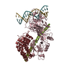 2f8xC  1ot8S S: Starting model for refinement C: citing same article ( |
|---|---|
| Similar structure data |


-Links
 PDBj
PDBj


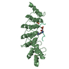
- Assembly
| Deposited unit | 
| ||||||||
|---|---|---|---|---|---|---|---|---|---|
| 1 | 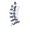
| ||||||||
| 2 | 
| ||||||||
| Unit cell |
|
-Components
| #1: Protein | Mass: 24502.348 Da / Num. of mol.: 2 / Fragment: ankyrin repeat domain, repeats 1-7 Source method: isolated from a genetically manipulated source Source: (gene. exp.) Homo sapiens (human) / Gene: Notch1 / Plasmid: pDEST15 / Production host:  Escherichia coli (E. coli) / Strain (production host): BL21(DE3)pLysS / References: UniProt: P46531 Escherichia coli (E. coli) / Strain (production host): BL21(DE3)pLysS / References: UniProt: P46531#2: Chemical | Sulfate  Mass: 96.063 Da / Num. of mol.: 2 / Source method: obtained synthetically / Formula: SO4 Mass: 96.063 Da / Num. of mol.: 2 / Source method: obtained synthetically / Formula: SO4#3: Water | ChemComp-HOH / | Water Mass: 18.015 Da / Num. of mol.: 404 / Source method: isolated from a natural source / Formula: H2O Mass: 18.015 Da / Num. of mol.: 404 / Source method: isolated from a natural source / Formula: H2O |
|---|
-Experimental details
-Experiment
| Experiment | Method: X-RAY DIFFRACTION / Number of used crystals: 1 |
|---|
- Sample preparation
| Crystal | Density Matthews: 3.18 Å3/Da / Density % sol: 61.34 % |
|---|
-Data collection
| Diffraction source | Source: SYNCHROTRON / Site: NSLS  / Beamline: X29A / Wavelength: 0.9791 Å / Beamline: X29A / Wavelength: 0.9791 Å |
|---|---|
| Detector | Type: ADSC QUANTUM 4 / Detector: CCD / Date: Jan 28, 2005 |
| Radiation | Protocol: SINGLE WAVELENGTH / Monochromatic (M) / Laue (L): M / Scattering type: x-ray |
| Radiation wavelength | Wavelength: 0.9791 Å / Relative weight: 1 |
| Reflection | Resolution: 1.55→30 Å / Num. all: 85821 / Num. obs: 70977 / % possible obs: 82.7 % / Observed criterion σ(F): 0 / Observed criterion σ(I): 0 / Redundancy: 2.2 % / Rsym value: 0.068 / Net I/σ(I): 11.9 |
| Reflection shell | Resolution: 1.55→1.61 Å / Redundancy: 2 % / Mean I/σ(I) obs: 2 / Num. unique all: 3220 / Rsym value: 0.375 / % possible all: 45.9 |
- Processing
| Software |
| ||||||||||||||||||||||||||||||||||||||||||||||||||||||||||||||||||||||||||||||||
|---|---|---|---|---|---|---|---|---|---|---|---|---|---|---|---|---|---|---|---|---|---|---|---|---|---|---|---|---|---|---|---|---|---|---|---|---|---|---|---|---|---|---|---|---|---|---|---|---|---|---|---|---|---|---|---|---|---|---|---|---|---|---|---|---|---|---|---|---|---|---|---|---|---|---|---|---|---|---|---|---|---|
| Refinement | Method to determine structure: MOLECULAR REPLACEMENT Starting model: PDB ENTRY 1OT8 Resolution: 1.55→30 Å / Rfactor Rfree error: 0.004 / Data cutoff high absF: 1901493.44 / Data cutoff low absF: 0 / Isotropic thermal model: RESTRAINED / Cross valid method: THROUGHOUT / σ(F): 0 / σ(I): 0 / Stereochemistry target values: Engh & Huber
| ||||||||||||||||||||||||||||||||||||||||||||||||||||||||||||||||||||||||||||||||
| Solvent computation | Solvent model: FLAT MODEL / Bsol: 71.4968 Å2 / ksol: 0.406579 e/Å3 | ||||||||||||||||||||||||||||||||||||||||||||||||||||||||||||||||||||||||||||||||
| Displacement parameters | Biso mean: 26.6 Å2
| ||||||||||||||||||||||||||||||||||||||||||||||||||||||||||||||||||||||||||||||||
| Refine analyze |
| ||||||||||||||||||||||||||||||||||||||||||||||||||||||||||||||||||||||||||||||||
| Refinement step | Cycle: LAST / Resolution: 1.55→30 Å
| ||||||||||||||||||||||||||||||||||||||||||||||||||||||||||||||||||||||||||||||||
| Refine LS restraints |
| ||||||||||||||||||||||||||||||||||||||||||||||||||||||||||||||||||||||||||||||||
| LS refinement shell | Resolution: 1.55→1.61 Å / Total num. of bins used: 6
| ||||||||||||||||||||||||||||||||||||||||||||||||||||||||||||||||||||||||||||||||
| Xplor file |
|
