 Movie
Movie Controller


+ Open data

- Basic information
| Entry | Database: PDB / ID: 2eg5 | ||||||
|---|---|---|---|---|---|---|---|
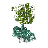
| Title | The structure of xanthosine methyltransferase | ||||||
 Components Components | Xanthosine methyltransferase | ||||||
 Keywords Keywords |  TRANSFERASE / SAM-dependant N-methyltransferase / xanthosine / SAH TRANSFERASE / SAM-dependant N-methyltransferase / xanthosine / SAH | ||||||
| Function / homology |  Function and homology information7-methylxanthosine synthase / alkaloid metabolic process / methyltransferase activity / methylation / metal ion binding Function and homology information7-methylxanthosine synthase / alkaloid metabolic process / methyltransferase activity / methylation / metal ion bindingSimilarity search - Function | ||||||
| Biological species |  Coffea canephora (robusta coffee) Coffea canephora (robusta coffee) | ||||||
| Method | X-RAY DIFFRACTION / SYNCHROTRON / MOLECULAR REPLACEMENT / Resolution: 2.2 Å | ||||||
 Authors Authors | McCarthy, A.A. / McCarthy, J.G. | ||||||
 Citation Citation | Journal: Plant Physiol. / Year: 2007 Title: The structure of two N-methyltransferases from the caffeine biosynthetic pathway Authors: McCarthy, A.A. / McCarthy, J.G. #1: Journal: Acta Crystallogr.,Sect.F / Year: 2007 Title: Cloning, expression, crystallization and preliminary X-ray analysis of the XMT and DXMT N-methyltransferases from Coffea canephora (robusta) Authors: McCarthy, A.A. / Biget, L. / Lin, C. / Petiard, V. / Tanksley, S.D. / McCarthy, J.G. | ||||||
| History |
|
- Structure visualization
| Structure viewer | Molecule: MolmilJmol/JSmol |
|---|
- Downloads & links
-Download
| PDBx/mmCIF format | 2eg5.cif.gz | 291.3 KB | Display | PDBx/mmCIF format |
|---|---|---|---|---|
| PDB format | pdb2eg5.ent.gz | 233.8 KB | Display | PDB format |
| PDBx/mmJSON format | 2eg5.json.gz | Tree view | PDBx/mmJSON format | |
| Others |  Other downloads Other downloads |
-Validation report
| Arichive directory | https://data.pdbj.org/pub/pdb/validation_reports/eg/2eg5ftp://data.pdbj.org/pub/pdb/validation_reports/eg/2eg5 | HTTPS FTP |
|---|
-Related structure data
| Related structure data | 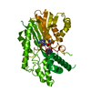 2efjSC S: Starting model for refinement C: citing same article ( |
|---|---|
| Similar structure data |


-Links
 PDBj
PDBj- Assembly
| Deposited unit | 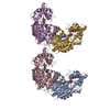
| ||||||||||||||||||||||||||||||||||||||||||||||||||||||||||||||||||||||||||||||||||||||||||||||||||||||||||||||||||||||||
|---|---|---|---|---|---|---|---|---|---|---|---|---|---|---|---|---|---|---|---|---|---|---|---|---|---|---|---|---|---|---|---|---|---|---|---|---|---|---|---|---|---|---|---|---|---|---|---|---|---|---|---|---|---|---|---|---|---|---|---|---|---|---|---|---|---|---|---|---|---|---|---|---|---|---|---|---|---|---|---|---|---|---|---|---|---|---|---|---|---|---|---|---|---|---|---|---|---|---|---|---|---|---|---|---|---|---|---|---|---|---|---|---|---|---|---|---|---|---|---|---|---|
| 1 | 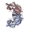
| ||||||||||||||||||||||||||||||||||||||||||||||||||||||||||||||||||||||||||||||||||||||||||||||||||||||||||||||||||||||||
| 2 | 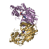
| ||||||||||||||||||||||||||||||||||||||||||||||||||||||||||||||||||||||||||||||||||||||||||||||||||||||||||||||||||||||||
| Unit cell |
| ||||||||||||||||||||||||||||||||||||||||||||||||||||||||||||||||||||||||||||||||||||||||||||||||||||||||||||||||||||||||
| Noncrystallographic symmetry (NCS) | NCS domain:
NCS domain segments: Component-ID: 1 / Refine code: 1
NCS ensembles :
| ||||||||||||||||||||||||||||||||||||||||||||||||||||||||||||||||||||||||||||||||||||||||||||||||||||||||||||||||||||||||
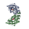
| Details | Chain A and C form a biological dimer / Chain E and G form a biological dimer |
-Components
| #1: Protein | Mass: 41894.137 Da / Num. of mol.: 4 Source method: isolated from a genetically manipulated source Source: (gene. exp.) Coffea canephora (robusta coffee) / Gene: XMT1 / Plasmid: pProEXHTb / Production host:  Escherichia coli (E. coli) / Strain (production host): BL21 (RIL) Escherichia coli (E. coli) / Strain (production host): BL21 (RIL)References: UniProt: A4GE69, Transferases; Transferring one-carbon groups; Methyltransferases#2: Chemical | ChemComp-SAH / S-Adenosyl-L-homocysteine  Type: L-peptide linking / Mass: 384.411 Da / Num. of mol.: 4 / Source method: obtained synthetically / Formula: C14H20N6O5S Type: L-peptide linking / Mass: 384.411 Da / Num. of mol.: 4 / Source method: obtained synthetically / Formula: C14H20N6O5S#3: Chemical | ChemComp-XTS / Xanthosine  Type: RNA linking / Mass: 284.225 Da / Num. of mol.: 4 / Source method: obtained synthetically / Formula: C10H12N4O6 Type: RNA linking / Mass: 284.225 Da / Num. of mol.: 4 / Source method: obtained synthetically / Formula: C10H12N4O6#4: Water | ChemComp-HOH / | Water Mass: 18.015 Da / Num. of mol.: 499 / Source method: isolated from a natural source / Formula: H2O Mass: 18.015 Da / Num. of mol.: 499 / Source method: isolated from a natural source / Formula: H2O |
|---|
-Experimental details
-Experiment
| Experiment | Method: X-RAY DIFFRACTION / Number of used crystals: 1 |
|---|
- Sample preparation
| Crystal | Density Matthews: 2.34 Å3/Da / Density % sol: 47.49 % |
|---|---|
| Crystal grow | Temperature: 293 K / Method: vapor diffusion, hanging drop / pH: 8.5 Details: 25% PEG 3350, 200mM Li2SO4, 100mM Tris-HCl, 1mM SAH, 1mM xanthosine, 2mM DTT, pH 8.5, VAPOR DIFFUSION, HANGING DROP, temperature 293K |
-Data collection
| Diffraction | Mean temperature: 100 K |
|---|---|
| Diffraction source | Source: SYNCHROTRON / Site: ESRF  / Beamline: ID14-4 / Wavelength: 0.9393 Å / Beamline: ID14-4 / Wavelength: 0.9393 Å |
| Detector | Type: ADSC QUANTUM 4 / Detector: CCD / Date: Nov 12, 2005 |
| Radiation | Monochromator: Khozu double crystal (Si 111) / Protocol: SINGLE WAVELENGTH / Monochromatic (M) / Laue (L): M / Scattering type: x-ray |
| Radiation wavelength | Wavelength: 0.9393 Å / Relative weight: 1 |
| Reflection | Resolution: 2.2→30 Å / Num. all: 75624 / Num. obs: 74901 / % possible obs: 95.2 % / Observed criterion σ(F): 0 / Observed criterion σ(I): 0 / Rmerge(I) obs: 0.079 / Net I/σ(I): 13.1 |
| Reflection shell | Resolution: 2.2→2.3 Å / Rmerge(I) obs: 0.443 / Mean I/σ(I) obs: 3 / Num. unique all: 10071 / % possible all: 93.4 |
- Processing
| Software |
| |||||||||||||||||||||||||||||||||||||||||||||||||||||||||||||||||||||||||||||||||||||||||||||||||||||||||||||||||||||||||||||
|---|---|---|---|---|---|---|---|---|---|---|---|---|---|---|---|---|---|---|---|---|---|---|---|---|---|---|---|---|---|---|---|---|---|---|---|---|---|---|---|---|---|---|---|---|---|---|---|---|---|---|---|---|---|---|---|---|---|---|---|---|---|---|---|---|---|---|---|---|---|---|---|---|---|---|---|---|---|---|---|---|---|---|---|---|---|---|---|---|---|---|---|---|---|---|---|---|---|---|---|---|---|---|---|---|---|---|---|---|---|---|---|---|---|---|---|---|---|---|---|---|---|---|---|---|---|---|
| Refinement | Method to determine structure: MOLECULAR REPLACEMENT Starting model: PDB ENTRY 2EFJ Resolution: 2.2→30 Å / Cor.coef. Fo:Fc: 0.933 / Cor.coef. Fo:Fc free: 0.896 / SU B: 17.957 / SU ML: 0.226 / TLS residual ADP flag: LIKELY RESIDUAL / Cross valid method: THROUGHOUT / ESU R: 0.345 / ESU R Free: 0.257 / Stereochemistry target values: MAXIMUM LIKELIHOOD / Details: HYDROGENS HAVE BEEN ADDED IN THE RIDING POSITIONS
| |||||||||||||||||||||||||||||||||||||||||||||||||||||||||||||||||||||||||||||||||||||||||||||||||||||||||||||||||||||||||||||
| Solvent computation | Ion probe radii: 0.8 Å / Shrinkage radii: 0.8 Å / VDW probe radii: 1.4 Å / Solvent model: BABINET MODEL WITH MASK | |||||||||||||||||||||||||||||||||||||||||||||||||||||||||||||||||||||||||||||||||||||||||||||||||||||||||||||||||||||||||||||
| Displacement parameters | Biso mean: 44.845 Å2
| |||||||||||||||||||||||||||||||||||||||||||||||||||||||||||||||||||||||||||||||||||||||||||||||||||||||||||||||||||||||||||||
| Refinement step | Cycle: LAST / Resolution: 2.2→30 Å
| |||||||||||||||||||||||||||||||||||||||||||||||||||||||||||||||||||||||||||||||||||||||||||||||||||||||||||||||||||||||||||||
| Refine LS restraints |
| |||||||||||||||||||||||||||||||||||||||||||||||||||||||||||||||||||||||||||||||||||||||||||||||||||||||||||||||||||||||||||||
| Refine LS restraints NCS | Refine-ID: X-RAY DIFFRACTION
| |||||||||||||||||||||||||||||||||||||||||||||||||||||||||||||||||||||||||||||||||||||||||||||||||||||||||||||||||||||||||||||
| LS refinement shell | Resolution: 2.2→2.257 Å / Total num. of bins used: 20
| |||||||||||||||||||||||||||||||||||||||||||||||||||||||||||||||||||||||||||||||||||||||||||||||||||||||||||||||||||||||||||||
| Refinement TLS params. | Method: refined / Refine-ID: X-RAY DIFFRACTION
| |||||||||||||||||||||||||||||||||||||||||||||||||||||||||||||||||||||||||||||||||||||||||||||||||||||||||||||||||||||||||||||
| Refinement TLS group |
|
