 Movie
Movie Controller

[English] 日本語
 Yorodumi
- PDB-2e6p: Solution structure of the Ig-like domain (714-804) from human Obs... -
+ Open data

- Basic information
| Entry | Database: PDB / ID: 2e6p | ||||||
|---|---|---|---|---|---|---|---|
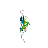
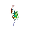
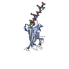
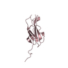
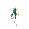
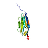
| Title | Solution structure of the Ig-like domain (714-804) from human Obscurin-like protein 1 | ||||||
 Components Components | Obscurin-like protein 1 | ||||||
 Keywords Keywords |  STRUCTURAL PROTEIN / Ig-like domain / Obscurin-like protein 1 / Structural Genomics / NPPSFA / National Project on Protein Structural and Functional Analyses / RIKEN Structural Genomics/Proteomics Initiative / RSGI STRUCTURAL PROTEIN / Ig-like domain / Obscurin-like protein 1 / Structural Genomics / NPPSFA / National Project on Protein Structural and Functional Analyses / RIKEN Structural Genomics/Proteomics Initiative / RSGI | ||||||
| Function / homology |  Function and homology information Function and homology information3M complex / protein localization to Golgi apparatus / cardiac myofibril assembly / cytoskeletal anchor activity / M band / positive regulation of dendrite morphogenesis / regulation of mitotic nuclear division / Golgi organization / intercalated disc / cytoskeleton organization ...3M complex / protein localization to Golgi apparatus / cardiac myofibril assembly / cytoskeletal anchor activity / M band / positive regulation of dendrite morphogenesis / regulation of mitotic nuclear division / Golgi organization / intercalated disc / cytoskeleton organization / microtubule cytoskeleton organization / Z disc / Neddylation / centrosome / perinuclear region of cytoplasm / Golgi apparatus / cytosol / cytoplasmSimilarity search - Function | ||||||
| Biological species |  Homo sapiens (human) Homo sapiens (human) | ||||||
| Method | SOLUTION NMR / Torsion angle dynamic | ||||||
 Authors Authors | Qin, X.R. / Suetake, T. / Hayashi, F. / Yokoyama, S. / RIKEN Structural Genomics/Proteomics Initiative (RSGI) | ||||||
 Citation Citation | Journal: To be published Title: Solution structure of the Ig-like domain (714-804) from human Obscurin-like protein 1 Authors: Qin, X.R. / Suetake, T. / Hayashi, F. / Yokoyama, S. | ||||||
| History |
|
- Structure visualization
| Structure viewer | Molecule: MolmilJmol/JSmol |
|---|
- Downloads & links
-Download
| PDBx/mmCIF format | 2e6p.cif.gz | 603.6 KB | Display | PDBx/mmCIF format |
|---|---|---|---|---|
| PDB format | pdb2e6p.ent.gz | 505.2 KB | Display | PDB format |
| PDBx/mmJSON format | 2e6p.json.gz | Tree view | PDBx/mmJSON format | |
| Others |  Other downloads Other downloads |
-Validation report
| Arichive directory | https://data.pdbj.org/pub/pdb/validation_reports/e6/2e6pftp://data.pdbj.org/pub/pdb/validation_reports/e6/2e6p | HTTPS FTP |
|---|
-Related structure data
| Similar structure data | |
|---|---|
| Other databases |


-Links
 PDBj
PDBj


- Assembly
| Deposited unit | 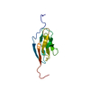
| |||||||||
|---|---|---|---|---|---|---|---|---|---|---|
| 1 |
| |||||||||
| NMR ensembles |
|
-Components
| #1: Protein | Mass: 11257.560 Da / Num. of mol.: 1 / Fragment: Ig-like domain Source method: isolated from a genetically manipulated source Source: (gene. exp.) Homo sapiens (human) / Description: cell free protein synthesis / Gene: OBSL1, KIAA0657 / Plasmid: P050919-02 / References: UniProt: O75147 |
|---|
-Experimental details
-Experiment
| Experiment | Method: SOLUTION NMR | ||||||||||||
|---|---|---|---|---|---|---|---|---|---|---|---|---|---|
| NMR experiment |
|
- Sample preparation
| Details | Contents: 1.17mM 13C, 15N-labeled protein; 20mM d-Tris-HCl(pH7.0); 100mM NaCl; 1mM d-DTT; 0.02% NaN3; 90% H2O, 10% D2O Solvent system: 90% H2O/10% D2O |
|---|---|
| Sample conditions | Ionic strength: 120mM / pH: 7.0 / Pressure: ambient / Temperature: 298 K |
-NMR measurement
| NMR spectrometer | Type: Varian INOVA / Manufacturer: Varian / Model: INOVA / Field strength: 800 MHz |
|---|
- Processing
| NMR software |
| ||||||||||||||||||||||||||||
|---|---|---|---|---|---|---|---|---|---|---|---|---|---|---|---|---|---|---|---|---|---|---|---|---|---|---|---|---|---|
| Refinement | Method: Torsion angle dynamic / Software ordinal: 1 | ||||||||||||||||||||||||||||
| NMR representative | Selection criteria: lowest energy | ||||||||||||||||||||||||||||
| NMR ensemble | Conformer selection criteria: structures with the least restraint violations, structures with the lowest energy, target function Conformers calculated total number: 100 / Conformers submitted total number: 20 |
