 Movie
Movie Controller


+ Open data

- Basic information
| Entry | Database: PDB / ID: 6yj0 | ||||||
|---|---|---|---|---|---|---|---|
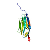
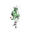
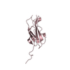
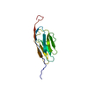
| Title | Solution NMR structure of titin N2A region Ig domain I83 | ||||||
 Components Components | Titin | ||||||
 Keywords Keywords | PROTEIN BINDING / calcium binding actin binding muscle I-band | ||||||
| Function / homology |  Function and homology information Function and homology informationforward locomotion / heart growth / striated muscle cell development / regulation of relaxation of cardiac muscle / sarcomerogenesis / structural molecule activity conferring elasticity / telethonin binding / skeletal muscle myosin thick filament assembly / cardiac myofibril assembly / muscle myosin complex ...forward locomotion / heart growth / striated muscle cell development / regulation of relaxation of cardiac muscle / sarcomerogenesis / structural molecule activity conferring elasticity / telethonin binding / skeletal muscle myosin thick filament assembly / cardiac myofibril assembly / muscle myosin complex / muscle alpha-actinin binding / ventricular system development / detection of muscle stretch / cardiac muscle tissue morphogenesis / adult heart development / cardiac muscle tissue development / cardiac muscle hypertrophy / M band / actinin binding / I band / cardiac muscle cell development / A band / structural constituent of muscle / ankyrin binding / sarcomere organization / skeletal muscle thin filament assembly / striated muscle thin filament / somitogenesis / heart morphogenesis / cardiac muscle contraction / protein kinase A signaling / sarcomere / condensed nuclear chromosome / muscle contraction / positive regulation of protein secretion / structural constituent of cytoskeleton / Z disc / response to calcium ion / : / actin filament binding / heart development / protein tyrosine kinase activity / in utero embryonic development / protease binding / calmodulin binding / non-specific serine/threonine protein kinase / phosphorylation / protein serine kinase activity / protein serine/threonine kinase activity / calcium ion binding / positive regulation of gene expression / protein kinase binding / enzyme binding / ATP binding / identical protein bindingSimilarity search - Function | ||||||
| Biological species |  Mus musculus (house mouse) Mus musculus (house mouse) | ||||||
| Method | SOLUTION NMR / simulated annealing | ||||||
 Authors Authors | Pfuhl, M. / Gage, M. | ||||||
 Citation Citation | Journal: J.Mol.Biol. / Year: 2021 Title: Solution NMR Structure of Titin N2A Region Ig Domain I83 and Its Interaction with Metal Ions. Authors: Kelly, C. / Pace, N. / Gage, M. / Pfuhl, M. | ||||||
| History |
|
- Structure visualization
| Structure viewer | Molecule: MolmilJmol/JSmol |
|---|
- Downloads & links
-Download
| PDBx/mmCIF format | 6yj0.cif.gz | 1.2 MB | Display | PDBx/mmCIF format |
|---|---|---|---|---|
| PDB format | pdb6yj0.ent.gz | 1 MB | Display | PDB format |
| PDBx/mmJSON format | 6yj0.json.gz | Tree view | PDBx/mmJSON format | |
| Others |  Other downloads Other downloads |
-Validation report
| Arichive directory | https://data.pdbj.org/pub/pdb/validation_reports/yj/6yj0ftp://data.pdbj.org/pub/pdb/validation_reports/yj/6yj0 | HTTPS FTP |
|---|
-Related structure data
| Similar structure data | |
|---|---|
| Other databases |


-Links
 PDBj
PDBj


- Assembly
| Deposited unit | 
| |||||||||
|---|---|---|---|---|---|---|---|---|---|---|
| 1 |
| |||||||||
| NMR ensembles |
|
-Components
| #1: Protein | / Connectin Mass: 11914.169 Da / Num. of mol.: 1 / Mutation: N.A. Source method: isolated from a genetically manipulated source Source: (gene. exp.) Mus musculus (house mouse) / Tissue: muscleSkeletal muscle / Cell: myocyte / Gene: Ttn / Variant: N2A / Plasmid: pET151/D-TOPO / Production host:  Escherichia coli BL21 (bacteria) / Variant (production host): BL21* Escherichia coli BL21 (bacteria) / Variant (production host): BL21*References: UniProt: A2ASS6, non-specific serine/threonine protein kinase |
|---|
-Experimental details
-Experiment
| Experiment | Method: SOLUTION NMR | ||||||||||||||||||||||||
|---|---|---|---|---|---|---|---|---|---|---|---|---|---|---|---|---|---|---|---|---|---|---|---|---|---|
| NMR experiment |
|
- Sample preparation
| Details |
| |||||||||||||||
|---|---|---|---|---|---|---|---|---|---|---|---|---|---|---|---|---|
| Sample |
| |||||||||||||||
| Sample conditions | Details: 20 mM Hepes, pH 7.2, 100 mM sodium chloride, 2 mM DTT and 0.02% NaN3 Ionic strength: 100 mM / Ionic strength err: 0.1 / Label: conditions_1 / pH: 7.2 / PH err: 0.02 / Pressure: 1 atm / Pressure err: 0.01 / Temperature: 298 K / Temperature err: 0.1 |
-NMR measurement
| NMR spectrometer |
|
|---|
- Processing
| NMR software |
| ||||||||||||||||
|---|---|---|---|---|---|---|---|---|---|---|---|---|---|---|---|---|---|
| Refinement | Method: simulated annealing / Software ordinal: 3 / Details: standard simulated annealing | ||||||||||||||||
| NMR representative | Selection criteria: lowest energy | ||||||||||||||||
| NMR ensemble | Conformer selection criteria: structures with the lowest energy Conformers calculated total number: 400 / Conformers submitted total number: 40 |
