 Movie
Movie Controller


+ Open data

- Basic information
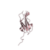
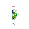
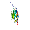
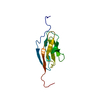
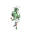
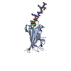
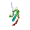
| Entry | Database: PDB / ID: 2dm2 | ||||||
|---|---|---|---|---|---|---|---|
| Title | Solution structure of the first ig domain of human palladin | ||||||
 Components Components | palladin | ||||||
 Keywords Keywords | PROTEIN BINDING / beta-sandwich / KIAA0992 / myopalladin / actin-associated scaffold / Structural Genomics / NPPSFA / National Project on Protein Structural and Functional Analyses / RIKEN Structural Genomics/Proteomics Initiative / RSGI | ||||||
| Function / homology |  Function and homology information Function and homology informationdendrite self-avoidance / cell-cell adhesion mediator activity / muscle alpha-actinin binding / epithelial cell morphogenesis / podosome / keratinocyte development / homophilic cell adhesion via plasma membrane adhesion molecules / excitatory synapse / stress fiber / cytoskeleton organization ...dendrite self-avoidance / cell-cell adhesion mediator activity / muscle alpha-actinin binding / epithelial cell morphogenesis / podosome / keratinocyte development / homophilic cell adhesion via plasma membrane adhesion molecules / excitatory synapse / stress fiber / cytoskeleton organization / ruffle / axon guidance / actin filament / Z disc / cell migration / actin cytoskeleton / lamellipodium / actin binding / growth cone / actin cytoskeleton organization / axon / focal adhesion / mitochondrion / nucleus / plasma membrane / cytosolSimilarity search - Function | ||||||
| Biological species |  Homo sapiens (human) Homo sapiens (human) | ||||||
| Method | SOLUTION NMR / torsion angle dynamics | ||||||
 Authors Authors | Nagashima, T. / Hayashi, F. / Yokoyama, S. / RIKEN Structural Genomics/Proteomics Initiative (RSGI) | ||||||
 Citation Citation | Journal: To be Published Title: Solution structure of the first ig domain of human palladin Authors: Nagashima, T. / Hayashi, F. / Yokoyama, S. | ||||||
| History |
|
- Structure visualization
| Structure viewer | Molecule: MolmilJmol/JSmol |
|---|
- Downloads & links
-Download
| PDBx/mmCIF format | 2dm2.cif.gz | 640.9 KB | Display | PDBx/mmCIF format |
|---|---|---|---|---|
| PDB format | pdb2dm2.ent.gz | 536.3 KB | Display | PDB format |
| PDBx/mmJSON format | 2dm2.json.gz | Tree view | PDBx/mmJSON format | |
| Others |  Other downloads Other downloads |
-Validation report
| Arichive directory | https://data.pdbj.org/pub/pdb/validation_reports/dm/2dm2ftp://data.pdbj.org/pub/pdb/validation_reports/dm/2dm2 | HTTPS FTP |
|---|
-Related structure data
| Similar structure data |
|---|


-Links
 PDBj
PDBj


- Assembly
| Deposited unit | 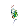
| |||||||||
|---|---|---|---|---|---|---|---|---|---|---|
| 1 |
| |||||||||
| NMR ensembles |
|
-Components
| #1: Antibody | Mass: 11923.438 Da / Num. of mol.: 1 / Fragment: IG Source method: isolated from a genetically manipulated source Source: (gene. exp.) Homo sapiens (human) / Description: Cell-free protein synthesis / Gene: KIAA0992 / Plasmid: P050711-06 / References: GenBank: 21361585, UniProt: Q8WX93*PLUS |
|---|
-Experimental details
-Experiment
| Experiment | Method: SOLUTION NMR | ||||||||||||
|---|---|---|---|---|---|---|---|---|---|---|---|---|---|
| NMR experiment |
|
- Sample preparation
| Details | Contents: 0.76mM uniformly 13C and 15N labeled protein; 20mM TrisHCl, 100mM NaCl, 1mM DTT, 0.02% NaN3, 10% D2O, 90% H2O Solvent system: 10% D2O, 90% H2O |
|---|---|
| Sample conditions | Ionic strength: 120mM / pH: 7.0 / Pressure: ambient / Temperature: 293 K |
-NMR measurement
| NMR spectrometer | Type: Varian INOVA / Manufacturer: Varian / Model: INOVA / Field strength: 800 MHz |
|---|
- Processing
| NMR software |
| ||||||||||||||||||||||||||||
|---|---|---|---|---|---|---|---|---|---|---|---|---|---|---|---|---|---|---|---|---|---|---|---|---|---|---|---|---|---|
| Refinement | Method: torsion angle dynamics / Software ordinal: 1 | ||||||||||||||||||||||||||||
| NMR representative | Selection criteria: lowest energy | ||||||||||||||||||||||||||||
| NMR ensemble | Conformer selection criteria: target function, structures with the lowest energy Conformers calculated total number: 100 / Conformers submitted total number: 20 |
