 Movie
Movie Controller

[English] 日本語
 Yorodumi
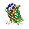
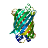
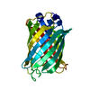
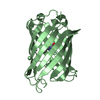
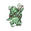
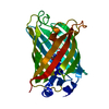
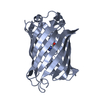
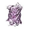
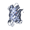
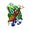
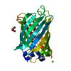
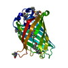
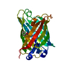
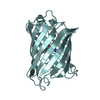
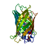
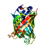
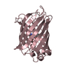
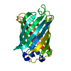
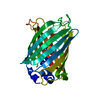
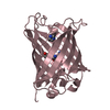
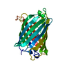
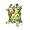
- PDB-2dug: crystal structure of a green fluorescent protein S65T/H148N at pH 5 -
+ Open data

- Basic information
| Entry | Database: PDB / ID: 2dug | ||||||
|---|---|---|---|---|---|---|---|
| Title | crystal structure of a green fluorescent protein S65T/H148N at pH 5 | ||||||
 Components Components | Green fluorescent protein | ||||||
 Keywords Keywords | LUMINESCENT PROTEIN / excited state proton transfer / very short hydrogen bond / green fluorescent protein | ||||||
| Function / homology |  Function and homology information Function and homology information | ||||||
| Biological species |   Aequorea victoria (jellyfish) Aequorea victoria (jellyfish) | ||||||
| Method | X-RAY DIFFRACTION / SYNCHROTRON / MOLECULAR REPLACEMENT / Resolution: 1.4 Å | ||||||
 Authors Authors | Shu, X. / Remington, S.J. | ||||||
 Citation Citation | Journal: Biochemistry / Year: 2007 Title: Ultrafast excited-state dynamics in the green fluorescent protein variant S65T/H148D. 1. Mutagenesis and structural studies. Authors: Shu, X. / Kallio, K. / Shi, X. / Abbyad, P. / Kanchanawong, P. / Childs, W. / Boxer, S.G. / Remington, S.J. | ||||||
| History |
|
- Structure visualization
| Structure viewer | Molecule: MolmilJmol/JSmol |
|---|
- Downloads & links
-Download
| PDBx/mmCIF format | 2dug.cif.gz | 58.6 KB | Display | PDBx/mmCIF format |
|---|---|---|---|---|
| PDB format | pdb2dug.ent.gz | 45.7 KB | Display | PDB format |
| PDBx/mmJSON format | 2dug.json.gz | Tree view | PDBx/mmJSON format | |
| Others |  Other downloads Other downloads |
-Validation report
| Arichive directory | https://data.pdbj.org/pub/pdb/validation_reports/du/2dugftp://data.pdbj.org/pub/pdb/validation_reports/du/2dug | HTTPS FTP |
|---|
-Related structure data


-Links
 PDBj
PDBj

- Assembly
| Deposited unit | 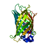
| ||||||||
|---|---|---|---|---|---|---|---|---|---|
| 1 |
| ||||||||
| Unit cell |
|
-Components
| #1: Protein | Mass: 26906.346 Da / Num. of mol.: 1 / Mutation: S65T, H148N, Q80R Source method: isolated from a genetically manipulated source Source: (gene. exp.) Aequorea victoria (jellyfish) / Production host:  Escherichia coli (E. coli) / References: UniProt: P42212 Escherichia coli (E. coli) / References: UniProt: P42212 |
|---|---|
| #2: Water | ChemComp-HOH / Water Mass: 18.015 Da / Num. of mol.: 233 / Source method: isolated from a natural source / Formula: H2O Mass: 18.015 Da / Num. of mol.: 233 / Source method: isolated from a natural source / Formula: H2O |
| Sequence details | RESIDUE SER 65 IS MUTATED TO THR 65. THR 65, TYR 66 AND GLY 67 ARE MODIFIED TO MAKE CHROMOPHOR |
-Experimental details
-Experiment
| Experiment | Method: X-RAY DIFFRACTION / Number of used crystals: 1 |
|---|
- Sample preparation
| Crystal | Density Matthews: 2.05 Å3/Da / Density % sol: 39.96 % |
|---|---|
| Crystal grow | Temperature: 298 K / Method: vapor diffusion, hanging drop / pH: 5 Details: 50mM Li2SO4, 100mM Acetate pH 9.5, 30% PEG 4000, VAPOR DIFFUSION, HANGING DROP, temperature 298K |
-Data collection
| Diffraction | Mean temperature: 100 K |
|---|---|
| Diffraction source | Source: SYNCHROTRON / Site: ALS  / Beamline: 8.2.2 / Wavelength: 1 Å / Beamline: 8.2.2 / Wavelength: 1 Å |
| Detector | Type: ADSC QUANTUM 315 / Detector: CCD / Date: Jan 15, 2006 |
| Radiation | Protocol: SINGLE WAVELENGTH / Monochromatic (M) / Laue (L): M / Scattering type: x-ray |
| Radiation wavelength | Wavelength: 1 Å / Relative weight: 1 |
| Reflection | Resolution: 1.4→50 Å / Num. all: 43567 / Num. obs: 43438 / % possible obs: 98.1 % / Observed criterion σ(F): 0 / Observed criterion σ(I): 0 |
| Reflection shell | Resolution: 1.4→1.45 Å / % possible all: 85.9 |
- Processing
| Software |
| |||||||||||||||
|---|---|---|---|---|---|---|---|---|---|---|---|---|---|---|---|---|
| Refinement | Method to determine structure: MOLECULAR REPLACEMENT / Resolution: 1.4→6 Å / σ(F): 0
| |||||||||||||||
| Refinement step | Cycle: LAST / Resolution: 1.4→6 Å
|
