 Movie
Movie Controller

[English] 日本語
 Yorodumi
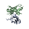
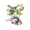
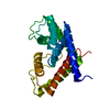
- PDB-2dud: Crystal structure of human mitochondrial single-stranded DNA-bind... -
+ Open data

- Basic information
| Entry | Database: PDB / ID: 2dud | ||||||
|---|---|---|---|---|---|---|---|
| Title | Crystal structure of human mitochondrial single-stranded DNA-binding protein(hmtSSB) | ||||||
 Components Components | Single-stranded DNA-binding protein | ||||||
 Keywords Keywords |  DNA BINDING PROTEIN / MITOCHONDRIA / SSB / Structural Genomics / NPPSFA / National Project on Protein Structural and Functional Analyses / RIKEN Structural Genomics/Proteomics Initiative / RSGI DNA BINDING PROTEIN / MITOCHONDRIA / SSB / Structural Genomics / NPPSFA / National Project on Protein Structural and Functional Analyses / RIKEN Structural Genomics/Proteomics Initiative / RSGI | ||||||
| Function / homology |  Function and homology information Function and homology informationpositive regulation of mitochondrial DNA replication / positive regulation of helicase activity / mitochondrial DNA replication / DNA unwinding involved in DNA replication / mitochondrial nucleoid / Transcriptional activation of mitochondrial biogenesis / single-stranded DNA binding / protein homotetramerization / mitochondrial matrix / chromatin binding ...positive regulation of mitochondrial DNA replication / positive regulation of helicase activity / mitochondrial DNA replication / DNA unwinding involved in DNA replication / mitochondrial nucleoid / Transcriptional activation of mitochondrial biogenesis / single-stranded DNA binding / protein homotetramerization / mitochondrial matrix / chromatin binding / protein homodimerization activity / mitochondrion / RNA binding / extracellular exosome / identical protein binding / nucleusSimilarity search - Function | ||||||
| Biological species |  Homo sapiens (human) Homo sapiens (human) | ||||||
| Method | X-RAY DIFFRACTION / SYNCHROTRON / MOLECULAR REPLACEMENT / Resolution: 2.7 Å | ||||||
 Authors Authors | Dong, X. / Bessho, Y. / Shirouzu, M. / Yokoyama, S. / RIKEN Structural Genomics/Proteomics Initiative (RSGI) | ||||||
 Citation Citation | Journal: To be Published Title: Crystal structure of human mitochondrial single-stranded DNA-binding protein(hmtSSB) Authors: Dong, X. / Bessho, Y. / Shirouzu, M. / Yokoyama, S. | ||||||
| History |
|
- Structure visualization
| Structure viewer | Molecule: MolmilJmol/JSmol |
|---|
- Downloads & links
-Download
| PDBx/mmCIF format | 2dud.cif.gz | 54.4 KB | Display | PDBx/mmCIF format |
|---|---|---|---|---|
| PDB format | pdb2dud.ent.gz | 38.5 KB | Display | PDB format |
| PDBx/mmJSON format | 2dud.json.gz | Tree view | PDBx/mmJSON format | |
| Others |  Other downloads Other downloads |
-Validation report
| Arichive directory | https://data.pdbj.org/pub/pdb/validation_reports/du/2dudftp://data.pdbj.org/pub/pdb/validation_reports/du/2dud | HTTPS FTP |
|---|
-Related structure data
| Related structure data |  1s3oS S: Starting model for refinement |
|---|---|
| Similar structure data | |
| Other databases |


-Links
 PDBj
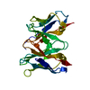
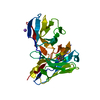
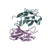
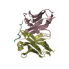
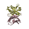
PDBj- Assembly
| Deposited unit | 
| ||||||||
|---|---|---|---|---|---|---|---|---|---|
| 1 |
| ||||||||
| 2 | 
| ||||||||
| Unit cell |
| ||||||||
| Details | The biological assembly is a dimer in the asymmetric |
-Components
| #1: Protein | Mass: 15354.269 Da / Num. of mol.: 2 / Fragment: Single-stranded DNA-binding protein, SSB Source method: isolated from a genetically manipulated source Source: (gene. exp.) Homo sapiens (human) / Description: cell-free protein synthesis / Plasmid: PX050223-18 / References: UniProt: Q04837 |
|---|
-Experimental details
-Experiment
| Experiment | Method: X-RAY DIFFRACTION / Number of used crystals: 1 |
|---|
- Sample preparation
| Crystal | Density Matthews: 2.42 Å3/Da / Density % sol: 49.24 % |
|---|---|
| Crystal grow | Temperature: 293 K / Method: vapor diffusion, sitting drop / pH: 9.5 Details: CHES, 0.2M NaCl, 10% PEG 8000, pH 9.5, VAPOR DIFFUSION, SITTING DROP, temperature 293K |
-Data collection
| Diffraction | Mean temperature: 100 K |
|---|---|
| Diffraction source | Source: SYNCHROTRON / Site: Photon Factory  / Beamline: BL-5A / Beamline: BL-5A |
| Detector | Type: ADSC QUANTUM 210 / Detector: CCD / Date: Apr 15, 2006 |
| Radiation | Monochromator: double flat Si (III) crystals / Protocol: SINGLE WAVELENGTH / Monochromatic (M) / Laue (L): M / Scattering type: x-ray |
| Radiation wavelength | Relative weight: 1 |
| Reflection | Resolution: 2.7→50 Å / Num. obs: 8832 / % possible obs: 100 % / Redundancy: 20.5 % / Biso Wilson estimate: 52.2 Å2 / Rsym value: 0.061 / Net I/σ(I): 68.4828 |
| Reflection shell | Resolution: 2.7→2.8 Å / Redundancy: 21.2 % / Mean I/σ(I) obs: 11.2 / Rsym value: 0.338 / % possible all: 100 |
- Processing
| Software |
| ||||||||||||||||||||||||||||||||||||||||||||||||||||||||||||||||||||||||||||||||
|---|---|---|---|---|---|---|---|---|---|---|---|---|---|---|---|---|---|---|---|---|---|---|---|---|---|---|---|---|---|---|---|---|---|---|---|---|---|---|---|---|---|---|---|---|---|---|---|---|---|---|---|---|---|---|---|---|---|---|---|---|---|---|---|---|---|---|---|---|---|---|---|---|---|---|---|---|---|---|---|---|---|
| Refinement | Method to determine structure: MOLECULAR REPLACEMENT Starting model: PDB ENTRY 1S3O Resolution: 2.7→34.99 Å / Rfactor Rfree error: 0.014 / Data cutoff high absF: 1599585.57 / Data cutoff low absF: 0 / Isotropic thermal model: RESTRAINED / Cross valid method: THROUGHOUT / σ(F): 0
| ||||||||||||||||||||||||||||||||||||||||||||||||||||||||||||||||||||||||||||||||
| Solvent computation | Solvent model: FLAT MODEL / Bsol: 38.8907 Å2 / ksol: 0.327667 e/Å3 | ||||||||||||||||||||||||||||||||||||||||||||||||||||||||||||||||||||||||||||||||
| Displacement parameters | Biso mean: 42.2 Å2
| ||||||||||||||||||||||||||||||||||||||||||||||||||||||||||||||||||||||||||||||||
| Refine analyze |
| ||||||||||||||||||||||||||||||||||||||||||||||||||||||||||||||||||||||||||||||||
| Refinement step | Cycle: LAST / Resolution: 2.7→34.99 Å
| ||||||||||||||||||||||||||||||||||||||||||||||||||||||||||||||||||||||||||||||||
| Refine LS restraints |
| ||||||||||||||||||||||||||||||||||||||||||||||||||||||||||||||||||||||||||||||||
| LS refinement shell | Resolution: 2.7→2.82 Å / Rfactor Rfree error: 0.052 / Total num. of bins used: 8
| ||||||||||||||||||||||||||||||||||||||||||||||||||||||||||||||||||||||||||||||||
| Xplor file |
|
