 Movie
Movie Controller


+ Open data

- Basic information
| Entry | Database: PDB / ID: 1s3o | ||||||
|---|---|---|---|---|---|---|---|
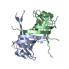
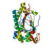
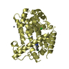
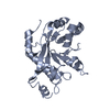
| Title | Human mitochondrial single strand DNA binding protein (hmSSB) | ||||||
 Components Components | Single-stranded DNA-binding protein, mitochondrial | ||||||
 Keywords Keywords |  DNA BINDING PROTEIN / OB fold DNA BINDING PROTEIN / OB fold | ||||||
| Function / homology |  Function and homology information Function and homology informationpositive regulation of mitochondrial DNA replication / positive regulation of helicase activity / mitochondrial DNA replication / DNA unwinding involved in DNA replication / mitochondrial nucleoid / Transcriptional activation of mitochondrial biogenesis / single-stranded DNA binding / protein homotetramerization / mitochondrial matrix / chromatin binding ...positive regulation of mitochondrial DNA replication / positive regulation of helicase activity / mitochondrial DNA replication / DNA unwinding involved in DNA replication / mitochondrial nucleoid / Transcriptional activation of mitochondrial biogenesis / single-stranded DNA binding / protein homotetramerization / mitochondrial matrix / chromatin binding / protein homodimerization activity / mitochondrion / RNA binding / extracellular exosome / identical protein binding / nucleusSimilarity search - Function | ||||||
| Biological species |  Homo sapiens (human) Homo sapiens (human) | ||||||
| Method | X-RAY DIFFRACTION / MIR / Resolution: 2.47 Å | ||||||
 Authors Authors | Venclovas, C. / Ginalski, K. / Kang, C. | ||||||
 Citation Citation | Journal: Protein Sci. / Year: 2004 Title: Sequence-structure mapping errors in the PDB: OB-fold domains Authors: Venclovas, C. / Ginalski, K. / Kang, C. | ||||||
| History |
|
- Structure visualization
| Structure viewer | Molecule: MolmilJmol/JSmol |
|---|
- Downloads & links
-Download
| PDBx/mmCIF format | 1s3o.cif.gz | 53.9 KB | Display | PDBx/mmCIF format |
|---|---|---|---|---|
| PDB format | pdb1s3o.ent.gz | 40.1 KB | Display | PDB format |
| PDBx/mmJSON format | 1s3o.json.gz | Tree view | PDBx/mmJSON format | |
| Others |  Other downloads Other downloads |
-Validation report
| Arichive directory | https://data.pdbj.org/pub/pdb/validation_reports/s3/1s3oftp://data.pdbj.org/pub/pdb/validation_reports/s3/1s3o | HTTPS FTP |
|---|
-Related structure data
| Related structure data | |
|---|---|
| Similar structure data |


-Links
 PDBj
PDBj- Assembly
| Deposited unit | 
| ||||||||
|---|---|---|---|---|---|---|---|---|---|
| 1 |
| ||||||||
| Unit cell |
|
-Components
| #1: Protein | Mass: 15216.124 Da / Num. of mol.: 2 / Source method: isolated from a natural source / Source: (natural) Homo sapiens (human) / References: UniProt: Q04837#2: Water | ChemComp-HOH / | Water Mass: 18.015 Da / Num. of mol.: 21 / Source method: isolated from a natural source / Formula: H2O Mass: 18.015 Da / Num. of mol.: 21 / Source method: isolated from a natural source / Formula: H2O |
|---|
-Experimental details
-Experiment
| Experiment | Method: X-RAY DIFFRACTION / Number of used crystals: 1 |
|---|
- Sample preparation
| Crystal | Density Matthews: 2.03 Å3/Da / Density % sol: 39.48 % |
|---|---|
| Crystal grow | Method: vapor diffusion, sitting drop / pH: 7 / Details: pH 7.0, VAPOR DIFFUSION, SITTING DROP |
-Data collection
| Diffraction | Mean temperature: 277 K |
|---|---|
| Diffraction source | Source: ROTATING ANODE |
| Detector | Detector: IMAGE PLATE |
| Radiation | Monochromator: yale mirror / Protocol: SINGLE WAVELENGTH / Monochromatic (M) / Laue (L): M / Scattering type: x-ray |
| Radiation wavelength | Relative weight: 1 |
| Reflection | Resolution: 2.4→25 Å / Num. all: 9790 / Num. obs: 9017 / % possible obs: 92.1 % / Observed criterion σ(F): 2 / Observed criterion σ(I): 2 |
- Processing
| Software |
| |||||||||||||||||||||
|---|---|---|---|---|---|---|---|---|---|---|---|---|---|---|---|---|---|---|---|---|---|---|
| Refinement | Method to determine structure: MIR / Resolution: 2.47→10 Å / σ(F): 2 / σ(I): 1.4
| |||||||||||||||||||||
| Refinement step | Cycle: LAST / Resolution: 2.47→10 Å
|
