 Movie
Movie Controller


+ Open data

- Basic information
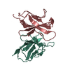
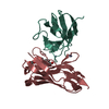
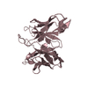
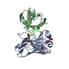
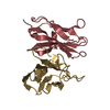
| Entry | Database: PDB / ID: 2otu | ||||||
|---|---|---|---|---|---|---|---|
| Title | Crystal structure of Fv polyglutamine complex | ||||||
 Components Components |
| ||||||
 Keywords Keywords |  IMMUNE SYSTEM / Antibody Fv polyglutamine complex IMMUNE SYSTEM / Antibody Fv polyglutamine complex | ||||||
| Function / homology |  Function and homology information Function and homology informationImmunoglobulin V-Type / Immunoglobulin V-set domain / Immunoglobulin V-set domain / Immunoglobulin subtype / Immunoglobulin / Ig-like domain profile. / Immunoglobulin-like domain / Immunoglobulin-like domain superfamily / Immunoglobulins / Immunoglobulin-like fold ...Immunoglobulin V-Type / Immunoglobulin V-set domain / Immunoglobulin V-set domain / Immunoglobulin subtype / Immunoglobulin / Ig-like domain profile. / Immunoglobulin-like domain / Immunoglobulin-like domain superfamily / Immunoglobulins / Immunoglobulin-like fold / Immunoglobulin-like / Sandwich / Mainly BetaSimilarity search - Domain/homology | ||||||
| Biological species |  Mus musculus (house mouse) Mus musculus (house mouse) | ||||||
| Method | X-RAY DIFFRACTION / SYNCHROTRON / MOLECULAR REPLACEMENT / Resolution: 1.68 Å | ||||||
 Authors Authors | Li, P. | ||||||
 Citation Citation | Journal: To be Published Title: Implications of the structure of a poly-Gln/anti-poly-Gln complex for disease progression and therapy Authors: Li, P. | ||||||
| History |
|
- Structure visualization
| Structure viewer | Molecule: MolmilJmol/JSmol |
|---|
- Downloads & links
-Download
| PDBx/mmCIF format | 2otu.cif.gz | 216.4 KB | Display | PDBx/mmCIF format |
|---|---|---|---|---|
| PDB format | pdb2otu.ent.gz | 173.7 KB | Display | PDB format |
| PDBx/mmJSON format | 2otu.json.gz | Tree view | PDBx/mmJSON format | |
| Others |  Other downloads Other downloads |
-Validation report
| Arichive directory | https://data.pdbj.org/pub/pdb/validation_reports/ot/2otuftp://data.pdbj.org/pub/pdb/validation_reports/ot/2otu | HTTPS FTP |
|---|
-Related structure data
| Related structure data |  2otwC  2gsgS S: Starting model for refinement C: citing same article ( |
|---|---|
| Similar structure data |


-Links
 PDBj
PDBj

- Assembly
| Deposited unit | 
| ||||||||
|---|---|---|---|---|---|---|---|---|---|
| 1 | 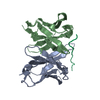
| ||||||||
| 2 | 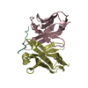
| ||||||||
| 3 | 
| ||||||||
| 4 | 
| ||||||||
| Unit cell |
|
-Components
| #1: Antibody | Mass: 12460.883 Da / Num. of mol.: 4 Source method: isolated from a genetically manipulated source Source: (gene. exp.) Mus musculus (house mouse) / Gene: VL / Plasmid: pET22b(+) / Species (production host): Escherichia coli / Production host:  Escherichia coli BL21(DE3) (bacteria) / Strain (production host): BL21(DE3) Escherichia coli BL21(DE3) (bacteria) / Strain (production host): BL21(DE3)#2: Antibody | Mass: 13330.936 Da / Num. of mol.: 4 Source method: isolated from a genetically manipulated source Source: (gene. exp.) Mus musculus (house mouse) / Gene: VH / Plasmid: pET22b(+) / Production host:   Spodoptera frugiperda (fall armyworm) / Strain (production host): BL21 (DE3) / References: UniProt: A2NN81 Spodoptera frugiperda (fall armyworm) / Strain (production host): BL21 (DE3) / References: UniProt: A2NN81#3: Protein/peptide | Mass: 1356.355 Da / Num. of mol.: 4 / Source method: obtained synthetically / Details: synthetic peptide #4: Water | ChemComp-HOH / | Water Mass: 18.015 Da / Num. of mol.: 928 / Source method: isolated from a natural source / Formula: H2O Mass: 18.015 Da / Num. of mol.: 928 / Source method: isolated from a natural source / Formula: H2O |
|---|
-Experimental details
-Experiment
| Experiment | Method: X-RAY DIFFRACTION / Number of used crystals: 1 |
|---|
- Sample preparation
| Crystal | Density Matthews: 2.48 Å3/Da / Density % sol: 50.33 % |
|---|---|
| Crystal grow | Temperature: 298 K / Method: evaporation / pH: 6 Details: 0.1 M sodium citrate, 0.2 M NH4Ac, 27.5% PEG 4000, pH 6.0, EVAPORATION, temperature 298K |
-Data collection
| Diffraction | Mean temperature: 173 K |
|---|---|
| Diffraction source | Source: SYNCHROTRON / Site: ALS  / Beamline: 8.2.2 / Wavelength: 1 / Beamline: 8.2.2 / Wavelength: 1 |
| Detector | Type: ADSC QUANTUM 315 / Detector: CCD / Date: Jan 1, 2006 / Details: Si(111) |
| Radiation | Monochromator: Si (111) / Protocol: SINGLE WAVELENGTH / Monochromatic (M) / Laue (L): M / Scattering type: x-ray |
| Radiation wavelength | Wavelength: 1 Å / Relative weight: 1 |
| Reflection | Resolution: 1.68→41 Å / Num. all: 119617 / Num. obs: 116627 / % possible obs: 97.5 % / Redundancy: 4.4 % / Rmerge(I) obs: 0.046 / Rsym value: 4.3 / Net I/σ(I): 34.3 |
| Reflection shell | Resolution: 1.68→1.74 Å / Redundancy: 3.5 % / Rmerge(I) obs: 0.258 / Mean I/σ(I) obs: 3.4 / Num. unique all: 10848 / Rsym value: 25.8 / % possible all: 91.1 |
- Processing
| Software |
| |||||||||||||||||||||||||
|---|---|---|---|---|---|---|---|---|---|---|---|---|---|---|---|---|---|---|---|---|---|---|---|---|---|---|
| Refinement | Method to determine structure: MOLECULAR REPLACEMENT Starting model: PDB entry 2GSG Resolution: 1.68→41 Å / Isotropic thermal model: isotropic / Cross valid method: THROUGHOUT / σ(F): 0 / σ(I): 0 / Stereochemistry target values: Engh & Huber
| |||||||||||||||||||||||||
| Refinement step | Cycle: LAST / Resolution: 1.68→41 Å
| |||||||||||||||||||||||||
| Refine LS restraints |
| |||||||||||||||||||||||||
| LS refinement shell | Highest resolution: 1.68 Å
|
