 Movie
Movie Controller

[English] 日本語
 Yorodumi
- PDB-2c7w: Crystal Structure of human vascular endothelial growth factor-B: ... -
+ Open data

- Basic information
| Entry | Database: PDB / ID: 2c7w | ||||||
|---|---|---|---|---|---|---|---|
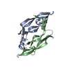
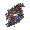
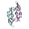
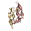
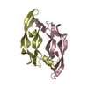
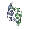
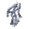
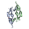
| Title | Crystal Structure of human vascular endothelial growth factor-B: Identification of amino acids important for angiogeninc activity | ||||||
 Components Components | VASCULAR ENDOTHELIAL GROWTH FACTOR B PRECURSOR | ||||||
 Keywords Keywords |  GROWTH FACTOR / VASCULAR ENDOTHELIAL GROWTH FACTOR-B / ANGIOGENESIS / CYSTEINE-KNOT MOTIF / TYROSINE KINASE / ISCHEMIA / MITOGEN / GLYCOPROTEIN / HEPARIN-BINDING GROWTH FACTOR / VASCULAR ENDOTHELIAL GROWTH FACTOR-B / ANGIOGENESIS / CYSTEINE-KNOT MOTIF / TYROSINE KINASE / ISCHEMIA / MITOGEN / GLYCOPROTEIN / HEPARIN-BINDING | ||||||
| Function / homology |  Function and homology information Function and homology informationpositive regulation of vascular wound healing / vascular endothelial growth factor receptor 1 binding / VEGF ligand-receptor interactions / positive regulation of mast cell chemotaxis / VEGF binds to VEGFR leading to receptor dimerization / induction of positive chemotaxis / vascular endothelial growth factor receptor 2 binding / protein O-linked glycosylation / coronary vasculature development / positive regulation of vascular endothelial growth factor receptor signaling pathway ...positive regulation of vascular wound healing / vascular endothelial growth factor receptor 1 binding / VEGF ligand-receptor interactions / positive regulation of mast cell chemotaxis / VEGF binds to VEGFR leading to receptor dimerization / induction of positive chemotaxis / vascular endothelial growth factor receptor 2 binding / protein O-linked glycosylation / coronary vasculature development / positive regulation of vascular endothelial growth factor receptor signaling pathway / sprouting angiogenesis / vascular endothelial growth factor signaling pathway / chemoattractant activity / positive regulation of cell division / vascular endothelial growth factor receptor signaling pathway / cardiac muscle contraction / positive regulation of endothelial cell proliferation / platelet alpha granule lumen / growth factor activity / positive regulation of angiogenesis / positive regulation of peptidyl-tyrosine phosphorylation / Platelet degranulation / heparin binding / negative regulation of neuron apoptotic process / positive regulation of phosphatidylinositol 3-kinase/protein kinase B signal transduction / positive regulation of ERK1 and ERK2 cascade / response to hypoxia / positive regulation of protein phosphorylation / negative regulation of gene expression / negative regulation of apoptotic process / extracellular space / extracellular region / membrane / identical protein bindingSimilarity search - Function | ||||||
| Biological species |  HOMO SAPIENS (human) HOMO SAPIENS (human) | ||||||
| Method | X-RAY DIFFRACTION / SYNCHROTRON / MOLECULAR REPLACEMENT / Resolution: 2.48 Å | ||||||
 Authors Authors | Iyer, S. / Scotney, P.D. / Nash, A.D. / Acharya, K.R. | ||||||
 Citation Citation | Journal: J.Mol.Biol. / Year: 2006 Title: Crystal Structure of Human Vascular Endothelial Growth Factor-B: Identification of Amino Acids Important for Receptor Binding Authors: Iyer, S. / Scotney, P.D. / Nash, A.D. / Acharya, K.R. | ||||||
| History |
| ||||||
| Remark 650 | HELIX DETERMINATION METHOD: AUTHOR PROVIDED. | ||||||
| Remark 700 | SHEET DETERMINATION METHOD: AUTHOR PROVIDED. |
- Structure visualization
| Structure viewer | Molecule: MolmilJmol/JSmol |
|---|
- Downloads & links
-Download
| PDBx/mmCIF format | 2c7w.cif.gz | 50.3 KB | Display | PDBx/mmCIF format |
|---|---|---|---|---|
| PDB format | pdb2c7w.ent.gz | 35.4 KB | Display | PDB format |
| PDBx/mmJSON format | 2c7w.json.gz | Tree view | PDBx/mmJSON format | |
| Others |  Other downloads Other downloads |
-Validation report
| Arichive directory | https://data.pdbj.org/pub/pdb/validation_reports/c7/2c7wftp://data.pdbj.org/pub/pdb/validation_reports/c7/2c7w | HTTPS FTP |
|---|
-Related structure data
| Related structure data | 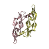 2vpfS S: Starting model for refinement |
|---|---|
| Similar structure data |
-Links
 PDBj
PDBj

- Assembly
| Deposited unit | 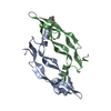
| ||||||||
|---|---|---|---|---|---|---|---|---|---|
| 1 |
| ||||||||
| Unit cell |
|
-Components
| #1: Protein | Mass: 11157.050 Da / Num. of mol.: 2 Source method: isolated from a genetically manipulated source Source: (gene. exp.) HOMO SAPIENS (human) / Plasmid: PET15B / Production host:  ESCHERICHIA COLI (E. coli) / References: UniProt: P49765 ESCHERICHIA COLI (E. coli) / References: UniProt: P49765#2: Chemical | ChemComp-MPD / ( | 2-Methyl-2,4-pentanediol  Mass: 118.174 Da / Num. of mol.: 1 / Source method: obtained synthetically / Formula: C6H14O2 / Comment: precipitant*YM Mass: 118.174 Da / Num. of mol.: 1 / Source method: obtained synthetically / Formula: C6H14O2 / Comment: precipitant*YM#3: Water | ChemComp-HOH / | Water Mass: 18.015 Da / Num. of mol.: 74 / Source method: isolated from a natural source / Formula: H2O Mass: 18.015 Da / Num. of mol.: 74 / Source method: isolated from a natural source / Formula: H2OCompound details | WORK AS A GROWTH FACTOR FOR ENDOTHELIA | |
|---|
-Experimental details
-Experiment
| Experiment | Method: X-RAY DIFFRACTION / Number of used crystals: 1 |
|---|
- Sample preparation
| Crystal | Density Matthews: 3.4 Å3/Da / Density % sol: 63.1 % |
|---|---|
| Crystal grow | pH: 6.8 Details: 0.1M HEPES (PH 6.8), 0.5M AMMONIUM SULPHATE 50%, MPD |
-Data collection
| Diffraction | Mean temperature: 100 K |
|---|---|
| Diffraction source | Source: SYNCHROTRON / Site: SRS  / Beamline: PX14.2 / Wavelength: 0.9795 / Beamline: PX14.2 / Wavelength: 0.9795 |
| Detector | Type: ADSC CCD / Detector: CCD / Date: Sep 18, 2003 / Details: MIRRORS |
| Radiation | Monochromator: SI 111 / Protocol: SINGLE WAVELENGTH / Monochromatic (M) / Laue (L): M / Scattering type: x-ray |
| Radiation wavelength | Wavelength: 0.9795 Å / Relative weight: 1 |
| Reflection | Resolution: 2.48→40 Å / Num. obs: 12054 / % possible obs: 97.4 % / Observed criterion σ(I): 0 / Redundancy: 9.1 % / Biso Wilson estimate: 43.1 Å2 / Rmerge(I) obs: 0.096 / Net I/σ(I): 12.9 |
| Reflection shell | Resolution: 2.48→2.57 Å / Redundancy: 10.1 % / Rmerge(I) obs: 0.63 / Mean I/σ(I) obs: 2.4 / % possible all: 99.3 |
- Processing
| Software |
| ||||||||||||||||||||||||||||||||||||||||||||||||||||||||||||
|---|---|---|---|---|---|---|---|---|---|---|---|---|---|---|---|---|---|---|---|---|---|---|---|---|---|---|---|---|---|---|---|---|---|---|---|---|---|---|---|---|---|---|---|---|---|---|---|---|---|---|---|---|---|---|---|---|---|---|---|---|---|
| Refinement | Method to determine structure: MOLECULAR REPLACEMENT Starting model: PDB ENTRY 2VPF Resolution: 2.48→40 Å / Rfactor Rfree error: 0.012 / Data cutoff high absF: 441769.31 / Isotropic thermal model: RESTRAINED / Cross valid method: THROUGHOUT / σ(F): 0 / Stereochemistry target values: MAXIMUM LIKELIHOOD
| ||||||||||||||||||||||||||||||||||||||||||||||||||||||||||||
| Solvent computation | Solvent model: FLAT MODEL / Bsol: 72.1563 Å2 / ksol: 0.379841 e/Å3 | ||||||||||||||||||||||||||||||||||||||||||||||||||||||||||||
| Displacement parameters | Biso mean: 40.2 Å2 | ||||||||||||||||||||||||||||||||||||||||||||||||||||||||||||
| Refine analyze |
| ||||||||||||||||||||||||||||||||||||||||||||||||||||||||||||
| Refinement step | Cycle: LAST / Resolution: 2.48→40 Å
| ||||||||||||||||||||||||||||||||||||||||||||||||||||||||||||
| Refine LS restraints |
| ||||||||||||||||||||||||||||||||||||||||||||||||||||||||||||
| LS refinement shell | Resolution: 2.48→2.57 Å / Rfactor Rwork: 0.312 / Total num. of bins used: 6 | ||||||||||||||||||||||||||||||||||||||||||||||||||||||||||||
| Xplor file |
|
