 Movie
Movie Controller


+ Open data

- Basic information
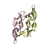
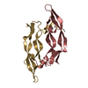
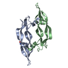
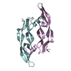
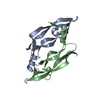
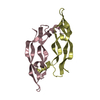
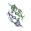
| Entry | Database: PDB / ID: 1vpf | ||||||
|---|---|---|---|---|---|---|---|
| Title | STRUCTURE OF HUMAN VASCULAR ENDOTHELIAL GROWTH FACTOR | ||||||
 Components Components | VASCULAR ENDOTHELIAL GROWTH FACTOR | ||||||
 Keywords Keywords | GROWTH FACTOR / CYSTINE KNOT / ANGIOGENESIS / VASCULOGENESIS | ||||||
| Function / homology |  Function and homology information Function and homology informationbasophil chemotaxis / cellular stress response to acid chemical / VEGF-A complex / Signaling by VEGF / lymph vessel morphogenesis / positive regulation of lymphangiogenesis / negative regulation of adherens junction organization / vascular endothelial growth factor receptor 1 binding / negative regulation of establishment of endothelial barrier / positive regulation of endothelial cell chemotaxis by VEGF-activated vascular endothelial growth factor receptor signaling pathway ...basophil chemotaxis / cellular stress response to acid chemical / VEGF-A complex / Signaling by VEGF / lymph vessel morphogenesis / positive regulation of lymphangiogenesis / negative regulation of adherens junction organization / vascular endothelial growth factor receptor 1 binding / negative regulation of establishment of endothelial barrier / positive regulation of endothelial cell chemotaxis by VEGF-activated vascular endothelial growth factor receptor signaling pathway / vascular endothelial growth factor receptor binding / VEGF ligand-receptor interactions / positive regulation of mast cell chemotaxis / post-embryonic camera-type eye development / primitive erythrocyte differentiation / positive regulation of protein kinase C signaling / positive regulation of cell proliferation by VEGF-activated platelet derived growth factor receptor signaling pathway / negative regulation of blood-brain barrier permeability / VEGF-activated neuropilin signaling pathway / bone trabecula formation / positive regulation of vascular endothelial growth factor signaling pathway / coronary vein morphogenesis / cardiac vascular smooth muscle cell development / lung vasculature development / lymphangiogenesis / eye photoreceptor cell development / positive regulation of trophoblast cell migration / endothelial cell chemotaxis / motor neuron migration / positive regulation of epithelial tube formation / vascular endothelial growth factor receptor-2 signaling pathway / VEGF binds to VEGFR leading to receptor dimerization / regulation of nitric oxide mediated signal transduction / positive regulation of axon extension involved in axon guidance / positive regulation of protein localization to early endosome / vascular wound healing / regulation of hematopoietic progenitor cell differentiation / positive regulation of blood vessel endothelial cell proliferation involved in sprouting angiogenesis / positive regulation of branching involved in ureteric bud morphogenesis / camera-type eye morphogenesis / neuropilin binding / induction of positive chemotaxis / coronary artery morphogenesis / negative regulation of cell-cell adhesion mediated by cadherin / vascular endothelial growth factor receptor 2 binding / dopaminergic neuron differentiation / negative regulation of epithelial to mesenchymal transition / tube formation / commissural neuron axon guidance / positive regulation of vascular permeability / platelet-derived growth factor receptor binding / surfactant homeostasis / extracellular matrix binding / cell migration involved in sprouting angiogenesis / retinal ganglion cell axon guidance / cardiac muscle cell development / epithelial cell maturation / sprouting angiogenesis / positive regulation of positive chemotaxis / endothelial cell proliferation / Regulation of gene expression by Hypoxia-inducible Factor / positive regulation of leukocyte migration / vascular endothelial growth factor signaling pathway / positive regulation of p38MAPK cascade / positive regulation of endothelial cell chemotaxis / artery morphogenesis / positive regulation of DNA biosynthetic process / branching involved in blood vessel morphogenesis / positive regulation of cell migration involved in sprouting angiogenesis / positive regulation of neuroblast proliferation / negative regulation of fat cell differentiation / positive chemotaxis / transmembrane receptor protein tyrosine kinase activator activity / positive regulation of sprouting angiogenesis / outflow tract morphogenesis / chemoattractant activity / positive regulation of focal adhesion assembly / mesoderm development / monocyte differentiation / positive regulation of receptor internalization / macrophage differentiation / positive regulation of cell division / fibronectin binding / positive regulation of cell adhesion / neuroblast proliferation / positive regulation of blood vessel endothelial cell migration / mammary gland alveolus development / cellular response to vascular endothelial growth factor stimulus / vasculogenesis / positive regulation of osteoblast differentiation / vascular endothelial growth factor receptor signaling pathway / heart morphogenesis / ovarian follicle development / cell maturation / homeostasis of number of cells within a tissue / positive regulation of protein autophosphorylation / positive regulation of endothelial cell proliferation / epithelial cell differentiation / lactation / TFAP2 (AP-2) family regulates transcription of growth factors and their receptorsSimilarity search - Function | ||||||
| Biological species |  Homo sapiens (human) Homo sapiens (human) | ||||||
| Method | X-RAY DIFFRACTION / MIR / Resolution: 2.5 Å | ||||||
 Authors Authors | Muller, Y.A. / De Vos, A.M. | ||||||
 Citation Citation | Journal: Proc.Natl.Acad.Sci.USA / Year: 1997 Title: Vascular endothelial growth factor: crystal structure and functional mapping of the kinase domain receptor binding site. Authors: Muller, Y.A. / Li, B. / Christinger, H.W. / Wells, J.A. / Cunningham, B.C. / de Vos, A.M. #1: Journal: Proteins / Year: 1996Title: Crystallization of the Receptor Binding Domain of Vascular Endothelial Growth Factor Authors: Christinger, H.W. / Muller, Y.A. / Berleau, L.T. / Keyt, B.A. / Cunningham, B.C. / Ferrara, N. / De Vos, A.M. | ||||||
| History |
|
- Structure visualization
| Structure viewer | Molecule: MolmilJmol/JSmol |
|---|
- Downloads & links
-Download
| PDBx/mmCIF format | 1vpf.cif.gz | 86 KB | Display | PDBx/mmCIF format |
|---|---|---|---|---|
| PDB format | pdb1vpf.ent.gz | 70.6 KB | Display | PDB format |
| PDBx/mmJSON format | 1vpf.json.gz | Tree view | PDBx/mmJSON format | |
| Others |  Other downloads Other downloads |
-Validation report
| Arichive directory | https://data.pdbj.org/pub/pdb/validation_reports/vp/1vpfftp://data.pdbj.org/pub/pdb/validation_reports/vp/1vpf | HTTPS FTP |
|---|
-Related structure data
| Similar structure data |
|---|
-Links
 PDBj
PDBj


- Assembly
| Deposited unit | 
| ||||||||||||||||
|---|---|---|---|---|---|---|---|---|---|---|---|---|---|---|---|---|---|
| 1 | 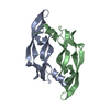
| ||||||||||||||||
| 2 | 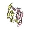
| ||||||||||||||||
| Unit cell |
| ||||||||||||||||
| Noncrystallographic symmetry (NCS) | NCS oper:
|
-Components
| #1: Protein | / VEGF / VASCULAR PERMEABILITY FACTOR / VPF Mass: 11948.680 Da / Num. of mol.: 4 / Fragment: RECEPTOR BINDING DOMAIN, RESIDUES 8 - 109 Source method: isolated from a genetically manipulated source Source: (gene. exp.) Homo sapiens (human) / Production host:  Escherichia coli (E. coli) / References: UniProt: P15692 Escherichia coli (E. coli) / References: UniProt: P15692#2: Water | ChemComp-HOH / | Water Mass: 18.015 Da / Num. of mol.: 191 / Source method: isolated from a natural source / Formula: H2O Mass: 18.015 Da / Num. of mol.: 191 / Source method: isolated from a natural source / Formula: H2O |
|---|
-Experimental details
-Experiment
| Experiment | Method: X-RAY DIFFRACTION / Number of used crystals: 1 |
|---|
- Sample preparation
| Crystal | Density Matthews: 2.7 Å3/Da / Density % sol: 54 % | ||||||||||||||||||||||||||||||||||||||||||||||||||||||
|---|---|---|---|---|---|---|---|---|---|---|---|---|---|---|---|---|---|---|---|---|---|---|---|---|---|---|---|---|---|---|---|---|---|---|---|---|---|---|---|---|---|---|---|---|---|---|---|---|---|---|---|---|---|---|---|
| Crystal grow | pH: 5.6 / Details: pH 5.6 | ||||||||||||||||||||||||||||||||||||||||||||||||||||||
| Crystal | *PLUS | ||||||||||||||||||||||||||||||||||||||||||||||||||||||
| Crystal grow | *PLUS pH: 7.5 / Method: vapor diffusion, sitting dropDetails: Christinger, H.W., (1996) Proteins: Struct.,Funct., Genet., 26, 353. | ||||||||||||||||||||||||||||||||||||||||||||||||||||||
| Components of the solutions | *PLUS
|
-Data collection
| Diffraction | Mean temperature: 100 K |
|---|---|
| Diffraction source | Source: ROTATING ANODE / Type: RIGAKU RUH2R / Wavelength: 1.5418 |
| Detector | Type: MARRESEARCH / Detector: IMAGE PLATE / Date: Dec 1, 1995 / Details: COLLIMATOR |
| Radiation | Monochromator: GRAPHITE(002) / Monochromatic (M) / Laue (L): M / Scattering type: x-ray |
| Radiation wavelength | Wavelength: 1.5418 Å / Relative weight: 1 |
| Reflection | Resolution: 2.5→10 Å / Num. obs: 17684 / % possible obs: 97 % / Redundancy: 3.6 % / Rsym value: 0.041 / Net I/σ(I): 56.5 |
| Reflection shell | Resolution: 2.5→2.6 Å / Redundancy: 3.2 % / Rsym value: 0.103 / % possible all: 96 |
| Reflection | *PLUS Rmerge(I) obs: 0.041 |
- Processing
| Software |
| ||||||||||||||||||||||||||||||||||||||||||||||||||||||||||||||||||||||||||||||||
|---|---|---|---|---|---|---|---|---|---|---|---|---|---|---|---|---|---|---|---|---|---|---|---|---|---|---|---|---|---|---|---|---|---|---|---|---|---|---|---|---|---|---|---|---|---|---|---|---|---|---|---|---|---|---|---|---|---|---|---|---|---|---|---|---|---|---|---|---|---|---|---|---|---|---|---|---|---|---|---|---|---|
| Refinement | Method to determine structure: MIR / Resolution: 2.5→10 Å / Data cutoff high absF: 100000 / Data cutoff low absF: 0.1 / Isotropic thermal model: RESTRAINED / Cross valid method: THROUGHOUT / σ(F): 0 Details: A SOLVENT MASK WAS APPLIED DURING THE FINAL REFINEMENT ROUNDS
| ||||||||||||||||||||||||||||||||||||||||||||||||||||||||||||||||||||||||||||||||
| Displacement parameters | Biso mean: 46.8 Å2
| ||||||||||||||||||||||||||||||||||||||||||||||||||||||||||||||||||||||||||||||||
| Refinement step | Cycle: LAST / Resolution: 2.5→10 Å
| ||||||||||||||||||||||||||||||||||||||||||||||||||||||||||||||||||||||||||||||||
| Refine LS restraints |
| ||||||||||||||||||||||||||||||||||||||||||||||||||||||||||||||||||||||||||||||||
| Refine LS restraints NCS | NCS model details: RESTRAINED | ||||||||||||||||||||||||||||||||||||||||||||||||||||||||||||||||||||||||||||||||
| LS refinement shell | Resolution: 2.5→2.59 Å / Total num. of bins used: 10
| ||||||||||||||||||||||||||||||||||||||||||||||||||||||||||||||||||||||||||||||||
| Xplor file |
| ||||||||||||||||||||||||||||||||||||||||||||||||||||||||||||||||||||||||||||||||
| Software | *PLUS Name: X-PLOR / Version: 3.8 / Classification: refinement | ||||||||||||||||||||||||||||||||||||||||||||||||||||||||||||||||||||||||||||||||
| Refine LS restraints | *PLUS
|
