 Movie
Movie Controller


+ Open data

- Basic information
| Entry | Database: PDB / ID: 2bh7 | ||||||
|---|---|---|---|---|---|---|---|
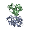
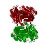
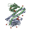
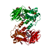
| Title | Crystal structure of a SeMet derivative of AmiD at 2.2 angstroms | ||||||
 Components Components | N-ACETYLMURAMOYL-L-ALANINE AMIDASE | ||||||
 Keywords Keywords | HYDROLASE / ZINC AMIDASE / PGRP / T7 LYSOZYME / AMPD | ||||||
| Function / homology |  Function and homology information Function and homology informationN-acetyl-anhydromuramoyl-L-alanine amidase activity / N-acetylmuramoyl-L-alanine amidase / peptidoglycan turnover / N-acetylmuramoyl-L-alanine amidase activity / outer membrane / peptidoglycan catabolic process / cell wall organization / cell outer membrane / zinc ion bindingSimilarity search - Function | ||||||
| Biological species |  ESCHERICHIA COLI (E. coli) ESCHERICHIA COLI (E. coli) | ||||||
| Method | X-RAY DIFFRACTION / SYNCHROTRON / SAD / Resolution: 2.2 Å | ||||||
 Authors Authors | Petrella, S. / Herman, R. / Sauvage, E. / Genereux, C. / Pennartz, A. / Joris, B. / Charlier, P. | ||||||
 Citation Citation | Journal: J.Mol.Biol. / Year: 2010 Title: Specific Structural Features of the N-Acetylmuramoyl-L-Alanine Amidase Amid from Escherichia Coli and Mechanistic Implications for Enzymes of This Family. Authors: Kerff, F. / Petrella, S. / Mercier, F. / Sauvage, E. / Herman, R. / Pennartz, A. / Zervosen, A. / Luxen, A. / Frere, J.M. / Joris, B. / Charlier, P. | ||||||
| History |
|
- Structure visualization
| Structure viewer | Molecule: MolmilJmol/JSmol |
|---|
- Downloads & links
-Download
| PDBx/mmCIF format | 2bh7.cif.gz | 63.8 KB | Display | PDBx/mmCIF format |
|---|---|---|---|---|
| PDB format | pdb2bh7.ent.gz | 51.4 KB | Display | PDB format |
| PDBx/mmJSON format | 2bh7.json.gz | Tree view | PDBx/mmJSON format | |
| Others |  Other downloads Other downloads |
-Validation report
| Arichive directory | https://data.pdbj.org/pub/pdb/validation_reports/bh/2bh7ftp://data.pdbj.org/pub/pdb/validation_reports/bh/2bh7 | HTTPS FTP |
|---|
-Related structure data


-Links
 PDBj
PDBj


- Assembly
| Deposited unit | 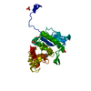
| ||||||||
|---|---|---|---|---|---|---|---|---|---|
| 1 | 
| ||||||||
| Unit cell |
|
-Components
| #1: Protein | Mass: 29563.814 Da / Num. of mol.: 1 / Source method: isolated from a natural source / Source: (natural) ESCHERICHIA COLI (E. coli) / Strain: K-12 MG1655References: UniProt: P75820, N-acetylmuramoyl-L-alanine amidase | ||||
|---|---|---|---|---|---|
| #2: Chemical | ChemComp-ZN /   Mass: 65.409 Da / Num. of mol.: 1 / Source method: obtained synthetically / Formula: Zn Mass: 65.409 Da / Num. of mol.: 1 / Source method: obtained synthetically / Formula: Zn | ||||
| #3: Chemical | ChemComp-SO4 / Sulfate  Mass: 96.063 Da / Num. of mol.: 6 / Source method: obtained synthetically / Formula: SO4 Mass: 96.063 Da / Num. of mol.: 6 / Source method: obtained synthetically / Formula: SO4#4: Water | ChemComp-HOH / | Water Mass: 18.015 Da / Num. of mol.: 147 / Source method: isolated from a natural source / Formula: H2O Mass: 18.015 Da / Num. of mol.: 147 / Source method: isolated from a natural source / Formula: H2OSequence details | RESIDUES 1 TO 17 WERE REMOVED AND A METHIONINE | |
-Experimental details
-Experiment
| Experiment | Method: X-RAY DIFFRACTION / Number of used crystals: 1 |
|---|
- Sample preparation
| Crystal | Density Matthews: 3.4 Å3/Da / Density % sol: 63 % |
|---|---|
| Crystal grow | pH: 4.6 / Details: 1M MGSO4 ACETATE PH 5.6 |
-Data collection
| Diffraction | Mean temperature: 100 K |
|---|---|
| Diffraction source | Source: SYNCHROTRON / Site: ESRF  / Beamline: BM30A / Wavelength: 0.9791 / Beamline: BM30A / Wavelength: 0.9791 |
| Detector | Type: MARRESEARCH / Detector: CCD / Date: Nov 6, 2004 |
| Radiation | Protocol: SINGLE WAVELENGTH / Monochromatic (M) / Laue (L): M / Scattering type: x-ray |
| Radiation wavelength | Wavelength: 0.9791 Å / Relative weight: 1 |
| Reflection | Resolution: 2.2→27.77 Å / Num. obs: 22663 / % possible obs: 99.8 % / Observed criterion σ(I): 2 / Redundancy: 48.3 % / Biso Wilson estimate: 27.9 Å2 / Rmerge(I) obs: 0.08 / Net I/σ(I): 57 |
| Reflection shell | Resolution: 2.2→2.32 Å / Redundancy: 23.1 % / Rmerge(I) obs: 0.4 / Mean I/σ(I) obs: 8.3 / % possible all: 99.8 |
- Processing
| Software |
| ||||||||||||||||||||||||||||||||||||||||||||||||||||||||||||||||||||||||||||||||
|---|---|---|---|---|---|---|---|---|---|---|---|---|---|---|---|---|---|---|---|---|---|---|---|---|---|---|---|---|---|---|---|---|---|---|---|---|---|---|---|---|---|---|---|---|---|---|---|---|---|---|---|---|---|---|---|---|---|---|---|---|---|---|---|---|---|---|---|---|---|---|---|---|---|---|---|---|---|---|---|---|---|
| Refinement | Method to determine structure: SAD / Resolution: 2.2→27.77 Å / Rfactor Rfree error: 0.006 / Data cutoff high absF: 2131583.74 / Isotropic thermal model: RESTRAINED / Cross valid method: THROUGHOUT / σ(F): 0
| ||||||||||||||||||||||||||||||||||||||||||||||||||||||||||||||||||||||||||||||||
| Solvent computation | Solvent model: FLAT MODEL / Bsol: 62.3704 Å2 / ksol: 0.388993 e/Å3 | ||||||||||||||||||||||||||||||||||||||||||||||||||||||||||||||||||||||||||||||||
| Displacement parameters | Biso mean: 36.4 Å2
| ||||||||||||||||||||||||||||||||||||||||||||||||||||||||||||||||||||||||||||||||
| Refine analyze |
| ||||||||||||||||||||||||||||||||||||||||||||||||||||||||||||||||||||||||||||||||
| Refinement step | Cycle: LAST / Resolution: 2.2→27.77 Å
| ||||||||||||||||||||||||||||||||||||||||||||||||||||||||||||||||||||||||||||||||
| Refine LS restraints |
| ||||||||||||||||||||||||||||||||||||||||||||||||||||||||||||||||||||||||||||||||
| LS refinement shell | Resolution: 2.2→2.34 Å / Rfactor Rfree error: 0.019 / Total num. of bins used: 6
| ||||||||||||||||||||||||||||||||||||||||||||||||||||||||||||||||||||||||||||||||
| Xplor file |
|
