 Movie
Movie Controller

[English] 日本語
 Yorodumi
- PDB-6w2z: Crystal Structure of the Beta Lactamase Class A PenP from Bacillu... -
+ Open data

- Basic information
| Entry | Database: PDB / ID: 6w2z | ||||||
|---|---|---|---|---|---|---|---|
| Title | Crystal Structure of the Beta Lactamase Class A PenP from Bacillus subtilis in the Complex with the Non-beta- lactam Beta-lactamase Inhibitor Avibactam | ||||||
 Components Components | Beta-lactamase | ||||||
 Keywords Keywords | HYDROLASE / beta lactamase class A / Inhibitor / Structural Genomics / Center for Structural Genomics of Infectious Diseases / CSGID | ||||||
| Function / homology |  Function and homology information Function and homology informationbeta-lactam antibiotic catabolic process / beta-lactamase activity / beta-lactamase / response to antibiotic / extracellular regionSimilarity search - Function | ||||||
| Biological species |  Bacillus subtilis (bacteria) Bacillus subtilis (bacteria) | ||||||
| Method | X-RAY DIFFRACTION / SYNCHROTRON / MOLECULAR REPLACEMENT / Resolution: 1.5 Å | ||||||
 Authors Authors | Kim, Y. / Maltseva, N. / Endres, M. / Joachimiak, A. / Center for Structural Genomics of Infectious Diseases (CSGID) | ||||||
| Funding support |  United States, 1items United States, 1items
| ||||||
 Citation Citation | Journal: To Be Published Title: Crystal Structure of the Beta Lactamase Class A PenP from Bacillus subtilis in the Complex with the Non-beta- lactam Beta-lactamase Inhibitor Avibactam Authors: Kim, Y. / Maltseva, N. / Endres, M. / Joachimiak, A. / Center for Structural Genomics of Infectious Diseases (CSGID) | ||||||
| History |
|
- Structure visualization
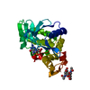
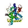
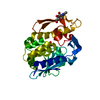
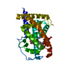
| Structure viewer | Molecule: MolmilJmol/JSmol |
|---|
- Downloads & links
-Download
| PDBx/mmCIF format | 6w2z.cif.gz | 253.5 KB | Display | PDBx/mmCIF format |
|---|---|---|---|---|
| PDB format | pdb6w2z.ent.gz | 179 KB | Display | PDB format |
| PDBx/mmJSON format | 6w2z.json.gz | Tree view | PDBx/mmJSON format | |
| Others |  Other downloads Other downloads |
-Validation report
| Arichive directory | https://data.pdbj.org/pub/pdb/validation_reports/w2/6w2zftp://data.pdbj.org/pub/pdb/validation_reports/w2/6w2z | HTTPS FTP |
|---|
-Related structure data
| Related structure data |  6ni1S S: Starting model for refinement |
|---|---|
| Similar structure data | |
| Other databases |


-Links
 PDBj
PDBj


- Assembly
| Deposited unit | 
| ||||||||||||
|---|---|---|---|---|---|---|---|---|---|---|---|---|---|
| 1 | 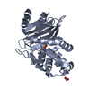
| ||||||||||||
| 2 | 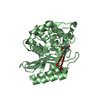
| ||||||||||||
| Unit cell |
|
-Components
| #1: Protein | / Penicillinase Mass: 30101.113 Da / Num. of mol.: 2 Source method: isolated from a genetically manipulated source Source: (gene. exp.) Bacillus subtilis (strain 168) (bacteria)Strain: 168 / Gene: penP, BSU18800 / Plasmid: pMCSG53 / Production host: Escherichia coli BL21(DE3) (bacteria) / Variant (production host): Gold / References: UniProt: P39824, beta-lactamase#2: Chemical | Avibactam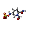  Mass: 267.260 Da / Num. of mol.: 2 / Source method: obtained synthetically / Formula: C7H13N3O6S / Feature type: SUBJECT OF INVESTIGATION / Comment: antibiotic, inhibitor*YM Mass: 267.260 Da / Num. of mol.: 2 / Source method: obtained synthetically / Formula: C7H13N3O6S / Feature type: SUBJECT OF INVESTIGATION / Comment: antibiotic, inhibitor*YM#3: Chemical | ChemComp-FMT / | Formic acid  Mass: 46.025 Da / Num. of mol.: 1 / Source method: obtained synthetically / Formula: CH2O2 Mass: 46.025 Da / Num. of mol.: 1 / Source method: obtained synthetically / Formula: CH2O2#4: Chemical | ChemComp-EDO / | Ethylene glycol  Mass: 62.068 Da / Num. of mol.: 1 / Source method: obtained synthetically / Formula: C2H6O2 Mass: 62.068 Da / Num. of mol.: 1 / Source method: obtained synthetically / Formula: C2H6O2#5: Water | ChemComp-HOH / | Water Mass: 18.015 Da / Num. of mol.: 352 / Source method: isolated from a natural source / Formula: H2O Mass: 18.015 Da / Num. of mol.: 352 / Source method: isolated from a natural source / Formula: H2OHas ligand of interest | Y | |
|---|
-Experimental details
-Experiment
| Experiment | Method: X-RAY DIFFRACTION / Number of used crystals: 1 |
|---|
- Sample preparation
| Crystal | Density Matthews: 1.96 Å3/Da / Density % sol: 37.18 % |
|---|---|
| Crystal grow | Temperature: 289 K / Method: vapor diffusion, sitting drop / pH: 8.8 Details: 50 mM Bicine pH 8.8, 2.9 %(v/v), 25.7 %(w/v) PEG2000 |
-Data collection
| Diffraction | Mean temperature: 100 K / Serial crystal experiment: N |
|---|---|
| Diffraction source | Source: SYNCHROTRON / Site: APS / Beamline: 19-BM / Wavelength: 0.97935 Å |
| Detector | Type: ADSC QUANTUM 210r / Detector: CCD / Date: Apr 10, 2019 |
| Radiation | Protocol: SINGLE WAVELENGTH / Monochromatic (M) / Laue (L): M / Scattering type: x-ray |
| Radiation wavelength | Wavelength: 0.97935 Å / Relative weight: 1 |
| Reflection | Resolution: 1.5→50 Å / Num. obs: 71543 / % possible obs: 95.4 % / Redundancy: 4.4 % / Biso Wilson estimate: 20.45 Å2 / Rmerge(I) obs: 0.06 / Net I/σ(I): 23.3 |
| Reflection shell | Resolution: 1.5→1.53 Å / Redundancy: 3.5 % / Rmerge(I) obs: 0.89 / Mean I/σ(I) obs: 1.41 / Num. unique obs: 3249 / CC1/2: 0.528 / % possible all: 86.3 |
- Processing
| Software |
| ||||||||||||||||||||||||||||||||||||||||||||||||||||||||||||||||||||||||||||||||||||||||||||||||||||||||||||||||||||||||||||||||||||||||||||||||||||||||||||||||||||||||||||||||||||||
|---|---|---|---|---|---|---|---|---|---|---|---|---|---|---|---|---|---|---|---|---|---|---|---|---|---|---|---|---|---|---|---|---|---|---|---|---|---|---|---|---|---|---|---|---|---|---|---|---|---|---|---|---|---|---|---|---|---|---|---|---|---|---|---|---|---|---|---|---|---|---|---|---|---|---|---|---|---|---|---|---|---|---|---|---|---|---|---|---|---|---|---|---|---|---|---|---|---|---|---|---|---|---|---|---|---|---|---|---|---|---|---|---|---|---|---|---|---|---|---|---|---|---|---|---|---|---|---|---|---|---|---|---|---|---|---|---|---|---|---|---|---|---|---|---|---|---|---|---|---|---|---|---|---|---|---|---|---|---|---|---|---|---|---|---|---|---|---|---|---|---|---|---|---|---|---|---|---|---|---|---|---|---|---|
| Refinement | Method to determine structure: MOLECULAR REPLACEMENT Starting model: PDBID 6NI1 Resolution: 1.5→27.1 Å / SU ML: 0.1782 / Cross valid method: FREE R-VALUE / σ(F): 1.38 / Phase error: 21.3077
| ||||||||||||||||||||||||||||||||||||||||||||||||||||||||||||||||||||||||||||||||||||||||||||||||||||||||||||||||||||||||||||||||||||||||||||||||||||||||||||||||||||||||||||||||||||||
| Solvent computation | Shrinkage radii: 0.9 Å / VDW probe radii: 1.11 Å | ||||||||||||||||||||||||||||||||||||||||||||||||||||||||||||||||||||||||||||||||||||||||||||||||||||||||||||||||||||||||||||||||||||||||||||||||||||||||||||||||||||||||||||||||||||||
| Displacement parameters | Biso mean: 25.77 Å2 | ||||||||||||||||||||||||||||||||||||||||||||||||||||||||||||||||||||||||||||||||||||||||||||||||||||||||||||||||||||||||||||||||||||||||||||||||||||||||||||||||||||||||||||||||||||||
| Refinement step | Cycle: LAST / Resolution: 1.5→27.1 Å
| ||||||||||||||||||||||||||||||||||||||||||||||||||||||||||||||||||||||||||||||||||||||||||||||||||||||||||||||||||||||||||||||||||||||||||||||||||||||||||||||||||||||||||||||||||||||
| Refine LS restraints |
| ||||||||||||||||||||||||||||||||||||||||||||||||||||||||||||||||||||||||||||||||||||||||||||||||||||||||||||||||||||||||||||||||||||||||||||||||||||||||||||||||||||||||||||||||||||||
| LS refinement shell |
|
