 Movie
Movie Controller

[English] 日本語
 Yorodumi
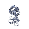
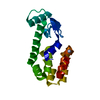
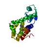
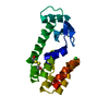
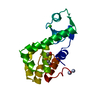
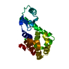
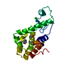
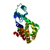
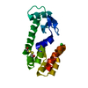
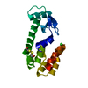
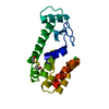
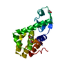
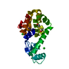
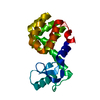
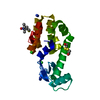
- PDB-2b7x: Sequential reorganization of beta-sheet topology by insertion of ... -
+ Open data

- Basic information
| Entry | Database: PDB / ID: 2b7x | ||||||
|---|---|---|---|---|---|---|---|
| Title | Sequential reorganization of beta-sheet topology by insertion of a single strand | ||||||
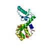
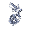
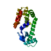
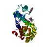
 Components Components | Lysozyme | ||||||
 Keywords Keywords | HYDROLASE / sequence duplication / protein design / structural switches / tandem repeat | ||||||
| Function / homology |  Function and homology information Function and homology informationviral release from host cell by cytolysis / peptidoglycan catabolic process / cell wall macromolecule catabolic process / lysozyme / lysozyme activity / host cell cytoplasm / defense response to bacteriumSimilarity search - Function | ||||||
| Biological species |  Enterobacteria phage T4 (virus) Enterobacteria phage T4 (virus) | ||||||
| Method | X-RAY DIFFRACTION / SYNCHROTRON / MOLECULAR REPLACEMENT / Resolution: 3 Å | ||||||
 Authors Authors | Sagermann, M. / Matthews, B.W. | ||||||
 Citation Citation | Journal: Protein Sci. / Year: 2006 Title: Sequential reorganization of beta-sheet topology by insertion of a single strand. Authors: Sagermann, M. / Baase, W.A. / Matthews, B.W. #1: Journal: Proc.Natl.Acad.Sci.USA / Year: 2003Title: Long distance conformation changes in a protein engineered by modulated sequence duplication Authors: Sagermann, M. / Gay, L. / Mattews, B.W. #2: Journal: Proc.Natl.Acad.Sci.USA / Year: 1999Title: Structural characterization of an engineered tandem repeat contrasts the importance of context and sequence in protein folding Authors: Sagermann, M. / Baase, W.A. / Matthews, B.W. | ||||||
| History |
|
- Structure visualization
| Structure viewer | Molecule: MolmilJmol/JSmol |
|---|
- Downloads & links
-Download
| PDBx/mmCIF format | 2b7x.cif.gz | 135.9 KB | Display | PDBx/mmCIF format |
|---|---|---|---|---|
| PDB format | pdb2b7x.ent.gz | 106.8 KB | Display | PDB format |
| PDBx/mmJSON format | 2b7x.json.gz | Tree view | PDBx/mmJSON format | |
| Others |  Other downloads Other downloads |
-Validation report
| Arichive directory | https://data.pdbj.org/pub/pdb/validation_reports/b7/2b7xftp://data.pdbj.org/pub/pdb/validation_reports/b7/2b7x | HTTPS FTP |
|---|
-Related structure data
| Related structure data | 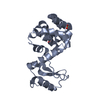 3jr6C 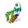 3lzmS C: citing same article ( S: Starting model for refinement |
|---|---|
| Similar structure data |


-Links
 PDBj
PDBj


- Assembly
| Deposited unit | 
| ||||||||
|---|---|---|---|---|---|---|---|---|---|
| 1 | 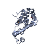
| ||||||||
| 2 | 
| ||||||||
| 3 | 
| ||||||||
| 4 | 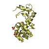
| ||||||||
| Unit cell |
|
-Components
| #1: Protein | / Lysis protein / Muramidase / Endolysin Mass: 19233.061 Da / Num. of mol.: 4 Mutation: C54T, C97A, residues (YTIGIG) inserted after residue G30 Source method: isolated from a genetically manipulated source Source: (gene. exp.) Enterobacteria phage T4 (virus) / Genus: T4-like viruses / Species: Enterobacteria phage T4 sensu lato / Gene: E / Plasmid: phs1403 / Production host:  Escherichia coli (E. coli) / Strain (production host): rr1 / References: UniProt: P00720, lysozyme Escherichia coli (E. coli) / Strain (production host): rr1 / References: UniProt: P00720, lysozyme#2: Chemical | ChemComp-SO4 / Sulfate  Mass: 96.063 Da / Num. of mol.: 8 / Source method: obtained synthetically / Formula: SO4 Mass: 96.063 Da / Num. of mol.: 8 / Source method: obtained synthetically / Formula: SO4 |
|---|
-Experimental details
-Experiment
| Experiment | Method: X-RAY DIFFRACTION / Number of used crystals: 1 |
|---|
- Sample preparation
| Crystal | Density Matthews: 2.23 Å3/Da / Density % sol: 44.9 % |
|---|---|
| Crystal grow | Temperature: 298 K / Method: vapor diffusion, hanging drop / pH: 7.8 Details: Polyehtylene glycol 4000, 50mM Ammonium sulfate, , pH 7.8, VAPOR DIFFUSION, HANGING DROP, temperature 298K |
-Data collection
| Diffraction | Mean temperature: 110 K |
|---|---|
| Diffraction source | Source: SYNCHROTRON / Site: ALS  / Beamline: 5.0.2 / Wavelength: 1 Å / Beamline: 5.0.2 / Wavelength: 1 Å |
| Detector | Type: ADSC QUANTUM 4 / Detector: CCD / Date: Nov 6, 1998 / Details: mirrors |
| Radiation | Monochromator: mirrors on si-crystal / Protocol: SINGLE WAVELENGTH / Monochromatic (M) / Laue (L): M / Scattering type: x-ray |
| Radiation wavelength | Wavelength: 1 Å / Relative weight: 1 |
| Reflection | Resolution: 3→20 Å / Num. all: 14371 / Num. obs: 10387 / % possible obs: 72.3 % / Observed criterion σ(F): 0 / Observed criterion σ(I): 0 / Rmerge(I) obs: 0.065 / Net I/σ(I): 7.4 |
| Reflection shell | Resolution: 3→3.25 Å / % possible all: 66.5 |
- Processing
| Software |
| ||||||||||||||||||||
|---|---|---|---|---|---|---|---|---|---|---|---|---|---|---|---|---|---|---|---|---|---|
| Refinement | Method to determine structure: MOLECULAR REPLACEMENT Starting model: pdb entry 3LZM, Resolution: 3→20 Å / Cross valid method: THROUGHOUT / σ(F): 0 / Stereochemistry target values: Engh & Huber Details: data was collected on very fragile, thin crystal plates resulting in low occupancy of reflections in data set. Several (non-isomorphous data sets were collected to further verify ...Details: data was collected on very fragile, thin crystal plates resulting in low occupancy of reflections in data set. Several (non-isomorphous data sets were collected to further verify interpretation. Refinement was carried out with the programs refmac and CNS.the sequence between residues 32 and 44 (new mutant numbering) were deleted from the model due to poor density. The c-terminal residues N169 and L170 (new mutant numbering) could not be identified in the density and were therefore omitted.
| ||||||||||||||||||||
| Displacement parameters |
| ||||||||||||||||||||
| Refinement step | Cycle: LAST / Resolution: 3→20 Å
| ||||||||||||||||||||
| Refine LS restraints |
|
