 Movie
Movie Controller


+ Open data

- Basic information
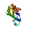
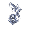
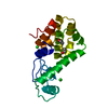
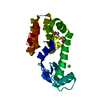
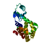
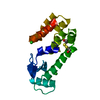
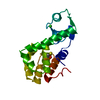
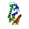
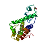
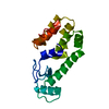
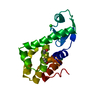
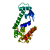
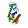
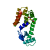
| Entry | Database: PDB / ID: 1p56 | ||||||
|---|---|---|---|---|---|---|---|
| Title | Duplication-extension of Helix A of T4 lysozyme | ||||||
 Components Components | PROTEIN (Lysozyme) | ||||||
 Keywords Keywords |  HYDROLASE / sequence duplication / folding propensity / completion folding experiment HYDROLASE / sequence duplication / folding propensity / completion folding experiment | ||||||
| Function / homology |  Function and homology information Function and homology informationviral release from host cell by cytolysis / peptidoglycan catabolic process / cell wall macromolecule catabolic process / lysozyme / lysozyme activity / host cell cytoplasm / defense response to bacteriumSimilarity search - Function | ||||||
| Biological species |  Enterobacteria phage T4 (virus) Enterobacteria phage T4 (virus) | ||||||
| Method | X-RAY DIFFRACTION / SYNCHROTRON / MOLECULAR REPLACEMENT / Resolution: 1.8 Å | ||||||
 Authors Authors | Sagermann, M. / Gay, L. / Baase, W.A. / Matthews, B.W. | ||||||
 Citation Citation | Journal: Biochemistry / Year: 2004 Title: Relocation or duplication of the helix A sequence of T4 lysozyme causes only modest changes in structure but can increase or decrease the rate of folding. Authors: Sagermann, M. / Baase, W.A. / Mooers, B.H. / Gay, L. / Matthews, B.W. #1: Journal: J.Mol.Biol. / Year: 2002Title: Crystal structures of a T4-lysozyme duplication-extension mutant demonstrates that the highly conserved beta-sheet region has low intrinsic folding propensity Authors: Sagermann, M. / Matthews, B.W. #2: Journal: J.Mol.Biol. / Year: 1987Title: Structure of bacteriophage T4 lysozyme refined at 1.7 A resolution Authors: Weaver, L.H. / Matthews, B.W. #3: Journal: Proc.Natl.Acad.Sci.USA / Year: 1999Title: Structural characterization of an engineered tandem repeat contrasts the importance of context and sequence in protein folding Authors: Sagermann, M. / Baase, W.A. / Matthews, B.W. | ||||||
| History |
| ||||||
| Remark 999 | SEQUENCE Residue Leu 164 was deleted and sequence SGGAMNIFEMLRIDE was appended to the C-terminus |
- Structure visualization
| Structure viewer | Molecule: MolmilJmol/JSmol |
|---|
- Downloads & links
-Download
| PDBx/mmCIF format | 1p56.cif.gz | 48.9 KB | Display | PDBx/mmCIF format |
|---|---|---|---|---|
| PDB format | pdb1p56.ent.gz | 33.7 KB | Display | PDB format |
| PDBx/mmJSON format | 1p56.json.gz | Tree view | PDBx/mmJSON format | |
| Others |  Other downloads Other downloads |
-Validation report
| Arichive directory | https://data.pdbj.org/pub/pdb/validation_reports/p5/1p56ftp://data.pdbj.org/pub/pdb/validation_reports/p5/1p56 | HTTPS FTP |
|---|
-Related structure data
| Related structure data | 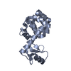 1p5cC 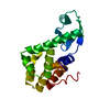 2lzmS S: Starting model for refinement C: citing same article ( |
|---|---|
| Similar structure data |


-Links
 PDBj
PDBj


- Assembly
| Deposited unit | 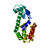
| ||||||||
|---|---|---|---|---|---|---|---|---|---|
| 1 |
| ||||||||
| Unit cell |
|
-Components
| #1: Protein | Mass: 19953.857 Da / Num. of mol.: 1 / Mutation: C54T, C97A Source method: isolated from a genetically manipulated source Source: (gene. exp.) Enterobacteria phage T4 (virus) / Genus: T4-like viruses / Species: Enterobacteria phage T4 sensu lato / Gene: Gene product E / Plasmid: phs1403 / Production host:  Escherichia coli (E. coli) / Strain (production host): RR1 / References: UniProt: P00720, lysozyme Escherichia coli (E. coli) / Strain (production host): RR1 / References: UniProt: P00720, lysozyme |
|---|---|
| #2: Water | ChemComp-HOH / Water Mass: 18.015 Da / Num. of mol.: 120 / Source method: isolated from a natural source / Formula: H2O Mass: 18.015 Da / Num. of mol.: 120 / Source method: isolated from a natural source / Formula: H2O |
-Experimental details
-Experiment
| Experiment | Method: X-RAY DIFFRACTION / Number of used crystals: 1 |
|---|
- Sample preparation
| Crystal | Density Matthews: 2.75 Å3/Da / Density % sol: 55.25 % |
|---|---|
| Crystal grow | Temperature: 298 K / Method: vapor diffusion, hanging drop / pH: 6.5 Details: 35% PEG 4000, 50mM phosphate buffer, 5% isopropanol, pH 6.5, VAPOR DIFFUSION, HANGING DROP, temperature 298K |
-Data collection
| Diffraction | Mean temperature: 170 K |
|---|---|
| Diffraction source | Source: SYNCHROTRON / Site: ALS  / Beamline: 5.0.2 / Wavelength: 0.999 Å / Beamline: 5.0.2 / Wavelength: 0.999 Å |
| Detector | Type: ADSC QUANTUM 4 / Detector: CCD / Date: Feb 10, 2002 / Details: mirrors |
| Radiation | Monochromator: Flat mirror, single SI crystal bend / Protocol: SINGLE WAVELENGTH / Monochromatic (M) / Laue (L): M / Scattering type: x-ray |
| Radiation wavelength | Wavelength: 0.999 Å / Relative weight: 1 |
| Reflection | Resolution: 1.7→28.75 Å / Num. obs: 22444 / % possible obs: 98.6 % / Observed criterion σ(F): 0 / Observed criterion σ(I): 0 / Redundancy: 3.6 % / Biso Wilson estimate: 14.24 Å2 / Rsym value: 0.066 / Net I/σ(I): 7.2 |
| Reflection shell | Resolution: 1.79→1.9 Å / Redundancy: 3.6 % / Mean I/σ(I) obs: 5.1 / Num. unique all: 3053 / Rsym value: 0.11 / % possible all: 98.7 |
- Processing
| Software |
| ||||||||||||||||||||
|---|---|---|---|---|---|---|---|---|---|---|---|---|---|---|---|---|---|---|---|---|---|
| Refinement | Method to determine structure: MOLECULAR REPLACEMENT Starting model: PDB ENTRY 2LZM Resolution: 1.8→30 Å / Isotropic thermal model: anisotropic / Cross valid method: THROUGHOUT / σ(F): 0 / Stereochemistry target values: Engh & Huber Details: Only disconnected density could be seen in the vicinity of the old c-terminus. Therefore, the sequence of the appended residues SGGAMNIFEMLRIDE could not be placed reliably into the density ...Details: Only disconnected density could be seen in the vicinity of the old c-terminus. Therefore, the sequence of the appended residues SGGAMNIFEMLRIDE could not be placed reliably into the density and was omitted in the final model.
| ||||||||||||||||||||
| Displacement parameters | Biso mean: 0.74 Å2
| ||||||||||||||||||||
| Refinement step | Cycle: LAST / Resolution: 1.8→30 Å
| ||||||||||||||||||||
| Refine LS restraints |
|
