 Movie
Movie Controller

[English] 日本語
 Yorodumi
- PDB-1jtm: Alternative Structures of a Sequence Extended T4 Lysozyme Show th... -
+ Open data

- Basic information
| Entry | Database: PDB / ID: 1jtm | ||||||
|---|---|---|---|---|---|---|---|
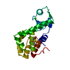
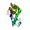
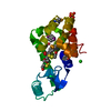
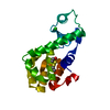
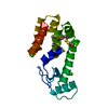
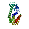
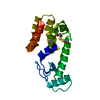
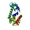
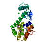
| Title | Alternative Structures of a Sequence Extended T4 Lysozyme Show that the Highly Conserved Beta-Sheet has Weak Intrinsic Folding Propensity | ||||||
 Components Components | LYSOZYME | ||||||
 Keywords Keywords | HYDROLASE / SEQUENCE DUPLICATION / CONTEXT DEPENDENT FOLDING / SEQUENCE REPEAT | ||||||
| Function / homology |  Function and homology information Function and homology informationviral release from host cell by cytolysis / peptidoglycan catabolic process / cell wall macromolecule catabolic process / lysozyme / lysozyme activity / host cell cytoplasm / defense response to bacteriumSimilarity search - Function | ||||||
| Biological species |  Enterobacteria phage T4 (virus) Enterobacteria phage T4 (virus) | ||||||
| Method | X-RAY DIFFRACTION / Resolution: 1.9 Å | ||||||
 Authors Authors | Sagermann, M. / Matthews, B.W. | ||||||
 Citation Citation | Journal: J.Mol.Biol. / Year: 2002 Title: Crystal Structures of a T4-lysozyme Duplication-extension Mutant Demonstrate that the Highly Conserved beta-Sheet Region has Low Intrinsic Folding Propensity Authors: Sagermann, M. / Matthews, B.W. #1: Journal: Proc.Natl.Acad.Sci.USA / Year: 1999Title: Structural Characterization of an Engineered Tandem Repeat contrasts the Importance of Context and Sequence in Protein Folding Authors: Sagermann, M. / Baase, W.A. / Matthews, B.W. #2: Journal: J.Mol.Biol. / Year: 1987Title: Structure of Bacteriophage T4 Lysozyme Refined at 1.7 A Resolution Authors: Weaver, L.H. / Matthews, B.W. | ||||||
| History |
|
- Structure visualization
| Structure viewer | Molecule: MolmilJmol/JSmol |
|---|
- Downloads & links
-Download
| PDBx/mmCIF format | 1jtm.cif.gz | 46.4 KB | Display | PDBx/mmCIF format |
|---|---|---|---|---|
| PDB format | pdb1jtm.ent.gz | 32.3 KB | Display | PDB format |
| PDBx/mmJSON format | 1jtm.json.gz | Tree view | PDBx/mmJSON format | |
| Others |  Other downloads Other downloads |
-Validation report
| Arichive directory | https://data.pdbj.org/pub/pdb/validation_reports/jt/1jtmftp://data.pdbj.org/pub/pdb/validation_reports/jt/1jtm | HTTPS FTP |
|---|
-Related structure data
| Related structure data |  1jtnC  2lzmS C: citing same article ( S: Starting model for refinement |
|---|---|
| Similar structure data |


-Links
 PDBj
PDBj


- Assembly
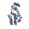
| Deposited unit | 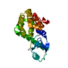
| ||||||||
|---|---|---|---|---|---|---|---|---|---|
| 1 |
| ||||||||
| Unit cell |
|
-Components
| #1: Protein | / LYSIS PROTEIN Mass: 20221.203 Da / Num. of mol.: 1 / Mutation: C54T, C97A Source method: isolated from a genetically manipulated source Source: (gene. exp.) Enterobacteria phage T4 (virus) / Genus: T4-like viruses / Species: Enterobacteria phage T4 sensu lato / Gene: E / Plasmid: PHS1403 / Production host:  Escherichia coli (E. coli) / Strain (production host): RR1 / References: UniProt: P00720, lysozyme Escherichia coli (E. coli) / Strain (production host): RR1 / References: UniProt: P00720, lysozyme |
|---|---|
| #2: Chemical | ChemComp-BME / 2-Mercaptoethanol  Mass: 78.133 Da / Num. of mol.: 1 / Source method: obtained synthetically / Formula: C2H6OS Mass: 78.133 Da / Num. of mol.: 1 / Source method: obtained synthetically / Formula: C2H6OS |
-Experimental details
-Experiment
| Experiment | Method: X-RAY DIFFRACTION / Number of used crystals: 1 |
|---|
- Sample preparation
| Crystal | Density Matthews: 2.75 Å3/Da / Density % sol: 55 % | |||||||||||||||||||||||||||||||||||||||||||||||||
|---|---|---|---|---|---|---|---|---|---|---|---|---|---|---|---|---|---|---|---|---|---|---|---|---|---|---|---|---|---|---|---|---|---|---|---|---|---|---|---|---|---|---|---|---|---|---|---|---|---|---|
| Crystal grow | Temperature: 298 K / Method: vapor diffusion, hanging drop / pH: 7 Details: 50MM TRIS-GLYCINE, 20% PEG 8000, 10% Isopropanol, pH 7.0, VAPOR DIFFUSION, HANGING DROP, temperature 298K | |||||||||||||||||||||||||||||||||||||||||||||||||
| Crystal grow | *PLUS pH: 7.5 / Details: used microseeding | |||||||||||||||||||||||||||||||||||||||||||||||||
| Components of the solutions | *PLUS
|
-Data collection
| Diffraction | Mean temperature: 170 K |
|---|---|
| Diffraction source | Source: ROTATING ANODE / Type: RIGAKU RU200 / Wavelength: 1.5418 Å |
| Detector | Type: RIGAKU RAXIS IV / Detector: IMAGE PLATE / Date: Jul 21, 1999 / Details: mirrors |
| Radiation | Monochromator: GRAPHITE / Protocol: SINGLE WAVELENGTH / Monochromatic (M) / Laue (L): M / Scattering type: x-ray |
| Radiation wavelength | Wavelength: 1.5418 Å / Relative weight: 1 |
| Reflection | Resolution: 1.9→17 Å / Num. all: 19736 / Num. obs: 19736 / % possible obs: 99.8 % / Observed criterion σ(F): 0 / Observed criterion σ(I): 0 / Redundancy: 4 % / Biso Wilson estimate: 24.7 Å2 / Rsym value: 0.069 / Net I/σ(I): 3.9 |
| Reflection shell | Resolution: 1.9→2.01 Å / Redundancy: 4 % / Mean I/σ(I) obs: 2.6 / Num. unique all: 2714 / Rsym value: 0.26 / % possible all: 99.8 |
| Reflection | *PLUS Rmerge(I) obs: 0.068 |
- Processing
| Software |
| ||||||||||||||||||||
|---|---|---|---|---|---|---|---|---|---|---|---|---|---|---|---|---|---|---|---|---|---|
| Refinement | Starting model: PDB ID 2LZM Resolution: 1.9→17 Å / Cross valid method: THROUGHOUT / σ(F): 0 / Stereochemistry target values: Engh & Huber Details: The structure was refined in the absence of water molecules. Only a BME molecule and a polyalanine peptide chain was fitted into difference density. The inserted polyalanine refined to a ...Details: The structure was refined in the absence of water molecules. Only a BME molecule and a polyalanine peptide chain was fitted into difference density. The inserted polyalanine refined to a grouped occupancy of ca. 0.3. Other less clear difference densities were observed as well but not fitted. The fitted chain was labeled with chain B. The connection of this chain to the c-terminus of the molecule is in very weak density and was not modeled.The former c-terminal residues Asn163 and Leu164 could were not detectable as well. Combination of CNS, BUSTER and TNT used for refinement.
| ||||||||||||||||||||
| Displacement parameters |
| ||||||||||||||||||||
| Refinement step | Cycle: LAST / Resolution: 1.9→17 Å
| ||||||||||||||||||||
| Refine LS restraints |
| ||||||||||||||||||||
| Refinement | *PLUS Highest resolution: 1.9 Å / Rfactor all: 0.219 / Rfactor obs: 0.214 / Rfactor Rfree: 0.265 / Rfactor Rwork: 0.214 | ||||||||||||||||||||
| Solvent computation | *PLUS | ||||||||||||||||||||
| Displacement parameters | *PLUS | ||||||||||||||||||||
| Refine LS restraints | *PLUS Type: t_angle_deg / Dev ideal: 1.4 |
