 Movie
Movie Controller

[English] 日本語
 Yorodumi
- PDB-2b7c: Yeast guanine nucleotide exchange factor eEF1Balpha K205A mutant ... -
+ Open data

- Basic information
| Entry | Database: PDB / ID: 2b7c | ||||||
|---|---|---|---|---|---|---|---|
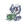
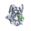
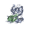
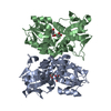
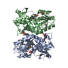
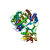
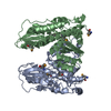
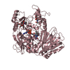
| Title | Yeast guanine nucleotide exchange factor eEF1Balpha K205A mutant in complex with eEF1A | ||||||
 Components Components |
| ||||||
 Keywords Keywords |  TRANSLATION / G-protein-GEF complex / eEF1A / eEF1Balpha TRANSLATION / G-protein-GEF complex / eEF1A / eEF1Balpha | ||||||
| Function / homology |  Function and homology information Function and homology informationEukaryotic Translation Elongation / eukaryotic translation elongation factor 1 complex / negative regulation of actin filament bundle assembly / HSF1 activation / melatonin binding / regulation of translational termination / tRNA export from nucleus / Protein methylation / fungal-type vacuole membrane / translational elongation ...Eukaryotic Translation Elongation / eukaryotic translation elongation factor 1 complex / negative regulation of actin filament bundle assembly / HSF1 activation / melatonin binding / regulation of translational termination / tRNA export from nucleus / Protein methylation / fungal-type vacuole membrane / translational elongation / actin filament bundle assembly / translation elongation factor activity / Neutrophil degranulation / cellular response to amino acid starvation / guanyl-nucleotide exchange factor activity / negative regulation of protein phosphorylation / maintenance of translational fidelity / negative regulation of protein kinase activity / GDP binding / actin filament binding / ribosome binding / cytoskeleton / ribosome / translation / GTPase activity / GTP binding / protein kinase binding / mitochondrion / cytosol / cytoplasmSimilarity search - Function | ||||||
| Biological species |  Saccharomyces cerevisiae (brewer's yeast) Saccharomyces cerevisiae (brewer's yeast) | ||||||
| Method | X-RAY DIFFRACTION / SYNCHROTRON / MOLECULAR REPLACEMENT / Resolution: 1.8 Å | ||||||
 Authors Authors | Pittman, Y.R. / Valente, L. / Jeppesen, M.G. / Andersen, G.R. / Patel, S. / Kinzy, T.G. | ||||||
 Citation Citation | Journal: J.Biol.Chem. / Year: 2006 Title: Mg2+ and a key lysine modulate exchange activity of eukaryotic translation elongation factor 1B alpha Authors: Pittman, Y.R. / Valente, L. / Jeppesen, M.G. / Andersen, G.R. / Patel, S. / Kinzy, T.G. #1: Journal: Mol.Cell / Year: 2000Title: Structural basis for nucleotide exchange and competition with tRNA in the yeast elongation factor complex eEF1A:eEF1Balpha Authors: Andersen, G.R. / Pedersen, L. / Valente, L. / Chatterjee, I. / Kinzy, T.G. / Kjeldgaard, M. / Nyborg, J. #2: Journal: Nat.Struct.Biol. / Year: 2001Title: Crystal structures of nucleotide exchange intermediates in the eEF1A-eEF1Balpha complex Authors: Andersen, G.R. / Valente, L. / Pedersen, L. / Kinzy, T.G. / Nyborg, J. | ||||||
| History |
|
- Structure visualization
| Structure viewer | Molecule: MolmilJmol/JSmol |
|---|
- Downloads & links
-Download
| PDBx/mmCIF format | 2b7c.cif.gz | 126.9 KB | Display | PDBx/mmCIF format |
|---|---|---|---|---|
| PDB format | pdb2b7c.ent.gz | 95.4 KB | Display | PDB format |
| PDBx/mmJSON format | 2b7c.json.gz | Tree view | PDBx/mmJSON format | |
| Others |  Other downloads Other downloads |
-Validation report
| Arichive directory | https://data.pdbj.org/pub/pdb/validation_reports/b7/2b7cftp://data.pdbj.org/pub/pdb/validation_reports/b7/2b7c | HTTPS FTP |
|---|
-Related structure data
| Related structure data |  2b7bC  1f60S S: Starting model for refinement C: citing same article ( |
|---|---|
| Similar structure data |


-Links
 PDBj
PDBj


- Assembly
| Deposited unit | 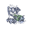
| ||||||||
|---|---|---|---|---|---|---|---|---|---|
| 1 |
| ||||||||
| Unit cell |
| ||||||||
| Details | The biological unit consists of one molecule each of eF1A and eEF1Balpha |
-Components
| #1: Protein | Mass: 50110.621 Da / Num. of mol.: 1 / Source method: isolated from a natural source / Source: (natural) Saccharomyces cerevisiae (brewer's yeast) / References: UniProt: P02994 |
|---|---|
| #2: Protein | Mass: 10414.727 Da / Num. of mol.: 1 / Fragment: C-terminal domain / Mutation: K1205A Source method: isolated from a genetically manipulated source Source: (gene. exp.) Saccharomyces cerevisiae (brewer's yeast)Gene: TEF5 / Plasmid: pET11d / Production host:  Escherichia coli (E. coli) / Strain (production host): BL211 / References: UniProt: P32471 Escherichia coli (E. coli) / Strain (production host): BL211 / References: UniProt: P32471 |
| #3: Water | ChemComp-HOH / Water Mass: 18.015 Da / Num. of mol.: 536 / Source method: isolated from a natural source / Formula: H2O Mass: 18.015 Da / Num. of mol.: 536 / Source method: isolated from a natural source / Formula: H2O |
-Experimental details
-Experiment
| Experiment | Method: X-RAY DIFFRACTION / Number of used crystals: 1 |
|---|
- Sample preparation
| Crystal | Density Matthews: 2.37 Å3/Da / Density % sol: 48.08 % |
|---|---|
| Crystal grow | Temperature: 277 K / Method: vapor diffusion, sitting drop / pH: 7.6 Details: 12% PEG 2000 MME, 100mM Tris-Cl pH 7.6, 100mM KCl, 3mM DTT, VAPOR DIFFUSION, SITTING DROP, temperature 277K |
-Data collection
| Diffraction | Mean temperature: 100 K |
|---|---|
| Diffraction source | Source: SYNCHROTRON / Site: ELETTRA  / Beamline: 5.2R / Wavelength: 0.9778 Å / Beamline: 5.2R / Wavelength: 0.9778 Å |
| Detector | Type: MARRESEARCH / Detector: CCD / Date: Feb 9, 2002 / Details: Three-segment Pt-coated toroidal mirrors |
| Radiation | Monochromator: Double Crystal (Si111, Si220) / Protocol: SINGLE WAVELENGTH / Monochromatic (M) / Laue (L): M / Scattering type: x-ray |
| Radiation wavelength | Wavelength: 0.9778 Å / Relative weight: 1 |
| Reflection | Resolution: 1.79→99 Å / Num. all: 47973 / Num. obs: 47973 / % possible obs: 88.6 % / Observed criterion σ(F): 0 / Observed criterion σ(I): -2 / Redundancy: 5.1 % / Rmerge(I) obs: 0.029 |
| Reflection shell | Resolution: 1.79→1.86 Å / Rmerge(I) obs: 0.231 / Mean I/σ(I) obs: 3.4 / % possible all: 69.9 |
- Processing
| Software |
| ||||||||||||||||||||
|---|---|---|---|---|---|---|---|---|---|---|---|---|---|---|---|---|---|---|---|---|---|
| Refinement | Method to determine structure: MOLECULAR REPLACEMENT Starting model: PBD entry 1F60 Resolution: 1.8→20 Å / Cross valid method: THROUGHOUT / σ(F): 0 / Stereochemistry target values: Engh & Huber
| ||||||||||||||||||||
| Refinement step | Cycle: LAST / Resolution: 1.8→20 Å
| ||||||||||||||||||||
| Refine LS restraints |
|
