 Movie
Movie Controller

[English] 日本語
 Yorodumi
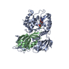
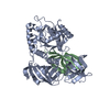
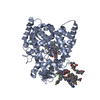
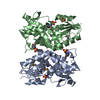
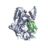
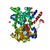
- PDB-1f60: CRYSTAL STRUCTURE OF THE YEAST ELONGATION FACTOR COMPLEX EEF1A:EEF1BA -
+ Open data

- Basic information
| Entry | Database: PDB / ID: 1f60 | ||||||
|---|---|---|---|---|---|---|---|
| Title | CRYSTAL STRUCTURE OF THE YEAST ELONGATION FACTOR COMPLEX EEF1A:EEF1BA | ||||||
 Components Components |
| ||||||
 Keywords Keywords |  TRANSLATION / protein-protein complex TRANSLATION / protein-protein complex | ||||||
| Function / homology |  Function and homology information Function and homology informationEukaryotic Translation Elongation / eukaryotic translation elongation factor 1 complex / negative regulation of actin filament bundle assembly / HSF1 activation / melatonin binding / regulation of translational termination / tRNA export from nucleus / Protein methylation / fungal-type vacuole membrane / translational elongation ...Eukaryotic Translation Elongation / eukaryotic translation elongation factor 1 complex / negative regulation of actin filament bundle assembly / HSF1 activation / melatonin binding / regulation of translational termination / tRNA export from nucleus / Protein methylation / fungal-type vacuole membrane / translational elongation / actin filament bundle assembly / translation elongation factor activity / Neutrophil degranulation / cellular response to amino acid starvation / guanyl-nucleotide exchange factor activity / negative regulation of protein phosphorylation / maintenance of translational fidelity / negative regulation of protein kinase activity / GDP binding / actin filament binding / ribosome binding / cytoskeleton / ribosome / translation / GTPase activity / GTP binding / protein kinase binding / mitochondrion / cytosol / cytoplasmSimilarity search - Function | ||||||
| Biological species |  Saccharomyces cerevisiae (brewer's yeast) Saccharomyces cerevisiae (brewer's yeast) | ||||||
| Method | X-RAY DIFFRACTION / SYNCHROTRON / Resolution: 1.67 Å | ||||||
 Authors Authors | Andersen, G.R. / Pedersen, L. / Valente, L. / Kinzy, T.G. / Nyborg, J. | ||||||
 Citation Citation | Journal: Mol.Cell / Year: 2000 Title: Structural basis for nucleotide exchange and competition with tRNA in the yeast elongation factor complex eEF1A:eEF1Balpha. Authors: Andersen, G.R. / Pedersen, L. / Valente, L. / Chatterjee, I. / Kinzy, T.G. / Kjeldgaard, M. / Nyborg, J. #1: Journal: Structure / Year: 1999Title: The Solution Structure of the Guanine Nucleotide Exchange Domain of Human Elongation Factor 1beta Reveals a Striking Resemblance to that of EF-Ts from Escherichia coli Authors: Perez, J.M. / Siegal, G. / Kriek, J. / Hard, K. / Dijk, J. / Canters, G.W. / Moeller, W. | ||||||
| History |
|
- Structure visualization
| Structure viewer | Molecule: MolmilJmol/JSmol |
|---|
- Downloads & links
-Download
| PDBx/mmCIF format | 1f60.cif.gz | 130.8 KB | Display | PDBx/mmCIF format |
|---|---|---|---|---|
| PDB format | pdb1f60.ent.gz | 99.3 KB | Display | PDB format |
| PDBx/mmJSON format | 1f60.json.gz | Tree view | PDBx/mmJSON format | |
| Others |  Other downloads Other downloads |
-Validation report
| Arichive directory | https://data.pdbj.org/pub/pdb/validation_reports/f6/1f60ftp://data.pdbj.org/pub/pdb/validation_reports/f6/1f60 | HTTPS FTP |
|---|
-Related structure data
| Related structure data | |
|---|---|
| Similar structure data |


-Links
 PDBj
PDBj


- Assembly
| Deposited unit | 
| ||||||||
|---|---|---|---|---|---|---|---|---|---|
| 1 |
| ||||||||
| Unit cell |
|
-Components
| #1: Protein | Mass: 50110.621 Da / Num. of mol.: 1 / Fragment: EEF1A / Source method: isolated from a natural source / Source: (natural) Saccharomyces cerevisiae (brewer's yeast) / References: UniProt: P02994 |
|---|---|
| #2: Protein | Mass: 10472.829 Da / Num. of mol.: 1 / Fragment: EEF1BA, CATALYTICAL C-TERMINAL DOMAIN Source method: isolated from a genetically manipulated source Source: (gene. exp.) Saccharomyces cerevisiae (brewer's yeast)Plasmid: PET11D / Production host:  Escherichia coli (E. coli) / References: UniProt: P32471 Escherichia coli (E. coli) / References: UniProt: P32471 |
| #3: Water | ChemComp-HOH / Water Mass: 18.015 Da / Num. of mol.: 733 / Source method: isolated from a natural source / Formula: H2O Mass: 18.015 Da / Num. of mol.: 733 / Source method: isolated from a natural source / Formula: H2O |
-Experimental details
-Experiment
| Experiment | Method: X-RAY DIFFRACTION / Number of used crystals: 1 |
|---|
- Sample preparation
| Crystal | Density Matthews: 2.25 Å3/Da / Density % sol: 45.29 % | ||||||||||||||||||||||||||||||||||||||||||||||||||||||
|---|---|---|---|---|---|---|---|---|---|---|---|---|---|---|---|---|---|---|---|---|---|---|---|---|---|---|---|---|---|---|---|---|---|---|---|---|---|---|---|---|---|---|---|---|---|---|---|---|---|---|---|---|---|---|---|
| Crystal grow | Temperature: 277 K / Method: vapor diffusion, sitting drop / pH: 8.5 Details: mmE 2000, Tris, Hepes, KCl, DTT, NaAzid, pH 8.5, VAPOR DIFFUSION, SITTING DROP, temperature 4K | ||||||||||||||||||||||||||||||||||||||||||||||||||||||
| Crystal grow | *PLUS Temperature: 277 K / pH: 7.2 Details: Pedersen, L.P., (2000) Acta Crystallogr., D57, 159. | ||||||||||||||||||||||||||||||||||||||||||||||||||||||
| Components of the solutions | *PLUS
|
-Data collection
| Diffraction | Mean temperature: 100 K |
|---|---|
| Diffraction source | Source: SYNCHROTRON / Site: EMBL/DESY, HAMBURG  / Beamline: BW7A / Wavelength: 0.9184 / Beamline: BW7A / Wavelength: 0.9184 |
| Detector | Type: MARRESEARCH / Detector: CCD / Date: Mar 1, 2000 |
| Radiation | Protocol: SINGLE WAVELENGTH / Monochromatic (M) / Laue (L): M / Scattering type: x-ray |
| Radiation wavelength | Wavelength: 0.9184 Å / Relative weight: 1 |
| Reflection | Resolution: 1.67→20 Å / Num. all: 65533 / Num. obs: 65533 / % possible obs: 99.3 % / Observed criterion σ(F): 0 / Observed criterion σ(I): 0 / Redundancy: 3.5 % / Biso Wilson estimate: 16.8 Å2 / Rmerge(I) obs: 0.04 / Net I/σ(I): 36.1 |
| Reflection shell | Resolution: 1.67→1.73 Å / Redundancy: 3 % / Rmerge(I) obs: 0.155 / Num. unique all: 5881 / % possible all: 93.5 |
| Reflection | *PLUS |
- Processing
| Software |
| |||||||||||||||||||||||||
|---|---|---|---|---|---|---|---|---|---|---|---|---|---|---|---|---|---|---|---|---|---|---|---|---|---|---|
| Refinement | Resolution: 1.67→20 Å / σ(F): 0 / σ(I): 0 Stereochemistry target values: engh & huber in arp_protin.doc Details: Refmac conjugate direction method
| |||||||||||||||||||||||||
| Refinement step | Cycle: LAST / Resolution: 1.67→20 Å
| |||||||||||||||||||||||||
| Refine LS restraints |
| |||||||||||||||||||||||||
| Software | *PLUS Name: REFMAC / Classification: refinement | |||||||||||||||||||||||||
| Refinement | *PLUS Lowest resolution: 20 Å / σ(F): 0 / Rfactor obs: 0.187 | |||||||||||||||||||||||||
| Solvent computation | *PLUS | |||||||||||||||||||||||||
| Displacement parameters | *PLUS | |||||||||||||||||||||||||
| Refine LS restraints | *PLUS Type: p_angle_d / Dev ideal: 1.6 |
