 Movie
Movie Controller

[English] 日本語
 Yorodumi
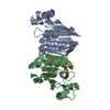
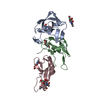
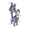
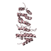
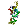
- PDB-2a25: Crystal structure of Siah1 SBD bound to the peptide EKPAAVVAPITTG... -
+ Open data

- Basic information
| Entry | Database: PDB / ID: 2a25 | ||||||
|---|---|---|---|---|---|---|---|
| Title | Crystal structure of Siah1 SBD bound to the peptide EKPAAVVAPITTG from SIP | ||||||
 Components Components |
| ||||||
 Keywords Keywords |  LIGASE / Protein-peptide complex LIGASE / Protein-peptide complex | ||||||
| Function / homology |  Function and homology information Function and homology informationNetrin-1 signaling / beta-catenin destruction complex / ubiquitin conjugating enzyme binding / S100 protein binding / SCF ubiquitin ligase complex / nuclear envelope lumen / anatomical structure morphogenesis / canonical Wnt signaling pathway / positive regulation of intrinsic apoptotic signaling pathway / tubulin binding ...Netrin-1 signaling / beta-catenin destruction complex / ubiquitin conjugating enzyme binding / S100 protein binding / SCF ubiquitin ligase complex / nuclear envelope lumen / anatomical structure morphogenesis / canonical Wnt signaling pathway / positive regulation of intrinsic apoptotic signaling pathway / tubulin binding / cellular response to leukemia inhibitory factor / axon guidance / RING-type E3 ubiquitin transferase / protein catabolic process / ubiquitin-protein transferase activity / ubiquitin protein ligase activity / Antigen processing: Ubiquitination & Proteasome degradation / nervous system development / heart development / ubiquitin-dependent protein catabolic process / spermatogenesis / proteasome-mediated ubiquitin-dependent protein catabolic process / neuron apoptotic process / amyloid fibril formation / protein ubiquitination / positive regulation of apoptotic process / cell cycle / Amyloid fiber formation / protein domain specific binding / apoptotic process / ubiquitin protein ligase binding / protein homodimerization activity / extracellular exosome / zinc ion binding / nucleoplasm / identical protein binding / nucleus / cytosol / cytoplasmSimilarity search - Function | ||||||
| Biological species |  Homo sapiens (human) Homo sapiens (human) | ||||||
| Method | X-RAY DIFFRACTION / MOLECULAR REPLACEMENT / Resolution: 2.2 Å | ||||||
 Authors Authors | Santelli, E. / Leone, M. / Li, C. / Fukushima, T. / Preece, N.E. / Olson, A.J. / Ely, K.R. / Reed, J.C. / Pellecchia, M. / Liddington, R.C. / Matsuzawa, S. | ||||||
 Citation Citation | Journal: J.Biol.Chem. / Year: 2005 Title: Structural Analysis of Siah1-Siah-interacting Protein Interactions and Insights into the Assembly of an E3 Ligase Multiprotein Complex Authors: Santelli, E. / Leone, M. / Li, C. / Fukushima, T. / Preece, N.E. / Olson, A.J. / Ely, K.R. / Reed, J.C. / Pellecchia, M. / Liddington, R.C. / Matsuzawa, S. | ||||||
| History |
|
- Structure visualization
| Structure viewer | Molecule: MolmilJmol/JSmol |
|---|
- Downloads & links
-Download
| PDBx/mmCIF format | 2a25.cif.gz | 49.1 KB | Display | PDBx/mmCIF format |
|---|---|---|---|---|
| PDB format | pdb2a25.ent.gz | 33.3 KB | Display | PDB format |
| PDBx/mmJSON format | 2a25.json.gz | Tree view | PDBx/mmJSON format | |
| Others |  Other downloads Other downloads |
-Validation report
| Arichive directory | https://data.pdbj.org/pub/pdb/validation_reports/a2/2a25ftp://data.pdbj.org/pub/pdb/validation_reports/a2/2a25 | HTTPS FTP |
|---|
-Related structure data
| Related structure data |  2a26C 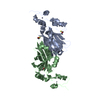 1k2fS S: Starting model for refinement C: citing same article ( |
|---|---|
| Similar structure data |


-Links
 PDBj
PDBj

- Assembly
| Deposited unit | 
| ||||||||
|---|---|---|---|---|---|---|---|---|---|
| 1 | 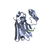
| ||||||||
| Unit cell |
| ||||||||
| Details | The second part of the biological assembly is generated by the two fold axis: 1-x, y, 1/2-z |
-Components
| #1: Protein | Mass: 21693.820 Da / Num. of mol.: 1 / Fragment: Substrate binding domain (residues 90-282) Source method: isolated from a genetically manipulated source Source: (gene. exp.) Homo sapiens (human) / Gene: SIAH1, HUMSIAH / Plasmid: pET15b / Species (production host): Escherichia coli / Production host:  Escherichia coli BL21(DE3) (bacteria) / Strain (production host): BL21(DE3) Escherichia coli BL21(DE3) (bacteria) / Strain (production host): BL21(DE3)References: UniProt: Q8IUQ4, Ligases; Forming carbon-nitrogen bonds; Acid-amino-acid ligases (peptide synthases) |
|---|---|
| #2: Protein/peptide | Mass: 1254.451 Da / Num. of mol.: 1 / Source method: obtained synthetically Details: The peptide was chemically synthesized. The sequence of the peptide can be naturally found in Homo sapiens (human) References: UniProt: Q9HB71 |
| #3: Chemical | ChemComp-ZN /   Mass: 65.409 Da / Num. of mol.: 1 / Source method: obtained synthetically / Formula: Zn Mass: 65.409 Da / Num. of mol.: 1 / Source method: obtained synthetically / Formula: Zn |
| #4: Water | ChemComp-HOH / Water Mass: 18.015 Da / Num. of mol.: 100 / Source method: isolated from a natural source / Formula: H2O Mass: 18.015 Da / Num. of mol.: 100 / Source method: isolated from a natural source / Formula: H2O |
-Experimental details
-Experiment
| Experiment | Method: X-RAY DIFFRACTION / Number of used crystals: 1 |
|---|
- Sample preparation
| Crystal | Density Matthews: 2.94 Å3/Da / Density % sol: 58 % |
|---|---|
| Crystal grow | Temperature: 293 K / Method: vapor diffusion, sitting drop / pH: 6 Details: sodium dihydrogen phospahte, di-potassium hydrogen phosphate, dithiothreithol, pH 6.0, VAPOR DIFFUSION, SITTING DROP, temperature 293K |
-Data collection
| Diffraction | Mean temperature: 100 K |
|---|---|
| Diffraction source | Source: ROTATING ANODE / Type: RIGAKU FR-E / Wavelength: 1.5418 Å |
| Detector | Type: RIGAKU RAXIS IV / Detector: IMAGE PLATE / Date: Jun 17, 2004 |
| Radiation | Protocol: SINGLE WAVELENGTH / Monochromatic (M) / Laue (L): M / Scattering type: x-ray |
| Radiation wavelength | Wavelength: 1.5418 Å / Relative weight: 1 |
| Reflection | Resolution: 2.2→50 Å / Num. obs: 10567 / Observed criterion σ(F): 0 / Observed criterion σ(I): -4 / Redundancy: 3.9 % / Biso Wilson estimate: 39.7 Å2 / Rmerge(I) obs: 0.043 / Net I/σ(I): 27 |
| Reflection shell | Resolution: 2.2→2.28 Å / Rmerge(I) obs: 0.365 / Mean I/σ(I) obs: 4 |
- Processing
| Software |
| ||||||||||||||||||||||||||||||||||||||||||||||||||||||||||||||||||||||||||||||||||||||||||||||||||||||||||||||||||||||||||||||||||||||||||||||||||||||||||||||||||||||||||
|---|---|---|---|---|---|---|---|---|---|---|---|---|---|---|---|---|---|---|---|---|---|---|---|---|---|---|---|---|---|---|---|---|---|---|---|---|---|---|---|---|---|---|---|---|---|---|---|---|---|---|---|---|---|---|---|---|---|---|---|---|---|---|---|---|---|---|---|---|---|---|---|---|---|---|---|---|---|---|---|---|---|---|---|---|---|---|---|---|---|---|---|---|---|---|---|---|---|---|---|---|---|---|---|---|---|---|---|---|---|---|---|---|---|---|---|---|---|---|---|---|---|---|---|---|---|---|---|---|---|---|---|---|---|---|---|---|---|---|---|---|---|---|---|---|---|---|---|---|---|---|---|---|---|---|---|---|---|---|---|---|---|---|---|---|---|---|---|---|---|---|---|
| Refinement | Method to determine structure: MOLECULAR REPLACEMENT Starting model: pdb entry 1k2f chain A Resolution: 2.2→29.11 Å / Cor.coef. Fo:Fc: 0.949 / Cor.coef. Fo:Fc free: 0.936 / SU B: 5.472 / SU ML: 0.14 / TLS residual ADP flag: LIKELY RESIDUAL / Cross valid method: THROUGHOUT / σ(F): 0 / ESU R: 0.253 / ESU R Free: 0.205 / Stereochemistry target values: MAXIMUM LIKELIHOOD / Details: HYDROGENS HAVE BEEN ADDED IN THE RIDING POSITIONS
| ||||||||||||||||||||||||||||||||||||||||||||||||||||||||||||||||||||||||||||||||||||||||||||||||||||||||||||||||||||||||||||||||||||||||||||||||||||||||||||||||||||||||||
| Solvent computation | Ion probe radii: 0.8 Å / Shrinkage radii: 0.8 Å / VDW probe radii: 1.4 Å / Solvent model: MASK | ||||||||||||||||||||||||||||||||||||||||||||||||||||||||||||||||||||||||||||||||||||||||||||||||||||||||||||||||||||||||||||||||||||||||||||||||||||||||||||||||||||||||||
| Displacement parameters | Biso mean: 26.77 Å2
| ||||||||||||||||||||||||||||||||||||||||||||||||||||||||||||||||||||||||||||||||||||||||||||||||||||||||||||||||||||||||||||||||||||||||||||||||||||||||||||||||||||||||||
| Refinement step | Cycle: LAST / Resolution: 2.2→29.11 Å
| ||||||||||||||||||||||||||||||||||||||||||||||||||||||||||||||||||||||||||||||||||||||||||||||||||||||||||||||||||||||||||||||||||||||||||||||||||||||||||||||||||||||||||
| Refine LS restraints |
| ||||||||||||||||||||||||||||||||||||||||||||||||||||||||||||||||||||||||||||||||||||||||||||||||||||||||||||||||||||||||||||||||||||||||||||||||||||||||||||||||||||||||||
| LS refinement shell | Resolution: 2.2→2.318 Å / Total num. of bins used: 10 /
| ||||||||||||||||||||||||||||||||||||||||||||||||||||||||||||||||||||||||||||||||||||||||||||||||||||||||||||||||||||||||||||||||||||||||||||||||||||||||||||||||||||||||||
| Refinement TLS params. | Method: refined / Origin x: 12.6082 Å / Origin y: 38.1424 Å / Origin z: 8.8587 Å
| ||||||||||||||||||||||||||||||||||||||||||||||||||||||||||||||||||||||||||||||||||||||||||||||||||||||||||||||||||||||||||||||||||||||||||||||||||||||||||||||||||||||||||
| Refinement TLS group |
|
