 Movie
Movie Controller

[English] 日本語
 Yorodumi
- PDB-3r4r: Crystal structure of a fimbrial assembly protein (BDI_3522) from ... -
+ Open data

- Basic information
| Entry | Database: PDB / ID: 3r4r | ||||||
|---|---|---|---|---|---|---|---|
| Title | Crystal structure of a fimbrial assembly protein (BDI_3522) from Parabacteroides distasonis ATCC 8503 at 2.38 A resolution | ||||||
 Components Components | hypothetical fimbrial assembly protein | ||||||
 Keywords Keywords |  CELL ADHESION / transthyretin-like (also known as prealbumin-like) / Structural Genomics / Joint Center for Structural Genomics / JCSG / Protein Structure Initiative / PSI-biology CELL ADHESION / transthyretin-like (also known as prealbumin-like) / Structural Genomics / Joint Center for Structural Genomics / JCSG / Protein Structure Initiative / PSI-biology | ||||||
| Function / homology | Immunoglobulin-like - #2590 / Immunoglobulin-like - #2580 / Major fimbrial subunit protein, N-terminal / Major fimbrial subunit protein (FimA) / pilus / Immunoglobulin-like / Sandwich / Mainly Beta / Putative fimbrium tip subunit Fim1F Function and homology information Function and homology information | ||||||
| Biological species |  Parabacteroides distasonis (bacteria) Parabacteroides distasonis (bacteria) | ||||||
| Method | X-RAY DIFFRACTION / SYNCHROTRON / MAD / Resolution: 2.38 Å | ||||||
 Authors Authors | Joint Center for Structural Genomics (JCSG) | ||||||
 Citation Citation | Journal: Cell / Year: 2016 Title: A Distinct Type of Pilus from the Human Microbiome. Authors: Xu, Q. / Shoji, M. / Shibata, S. / Naito, M. / Sato, K. / Elsliger, M.A. / Grant, J.C. / Axelrod, H.L. / Chiu, H.J. / Farr, C.L. / Jaroszewski, L. / Knuth, M.W. / Deacon, A.M. / Godzik, A. / ...Authors: Xu, Q. / Shoji, M. / Shibata, S. / Naito, M. / Sato, K. / Elsliger, M.A. / Grant, J.C. / Axelrod, H.L. / Chiu, H.J. / Farr, C.L. / Jaroszewski, L. / Knuth, M.W. / Deacon, A.M. / Godzik, A. / Lesley, S.A. / Curtis, M.A. / Nakayama, K. / Wilson, I.A. | ||||||
| History |
|
- Structure visualization
| Structure viewer | Molecule: MolmilJmol/JSmol |
|---|
- Downloads & links
-Download
| PDBx/mmCIF format | 3r4r.cif.gz | 221.2 KB | Display | PDBx/mmCIF format |
|---|---|---|---|---|
| PDB format | pdb3r4r.ent.gz | 180.7 KB | Display | PDB format |
| PDBx/mmJSON format | 3r4r.json.gz | Tree view | PDBx/mmJSON format | |
| Others |  Other downloads Other downloads |
-Validation report
| Arichive directory | https://data.pdbj.org/pub/pdb/validation_reports/r4/3r4rftp://data.pdbj.org/pub/pdb/validation_reports/r4/3r4r | HTTPS FTP |
|---|
-Related structure data
| Related structure data |  3liuC  3payC  3sy6C  3t2lC 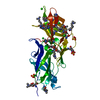 3ufiC  3up6C 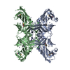 4dguC  4epsC 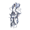 4gpvC  4h40C  4jg5C  4jrfC  4k4kC  4q98C  4qb7C  4qdgC  4rdbC  5cagC C: citing same article ( |
|---|---|
| Similar structure data | |
| Other databases |


-Links
 PDBj
PDBj- Assembly
| Deposited unit | 
| ||||||||
|---|---|---|---|---|---|---|---|---|---|
| 1 | 
| ||||||||
| 2 | 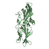
| ||||||||
| Unit cell |
|
-Components
| #1: Protein | Mass: 32903.355 Da / Num. of mol.: 2 Source method: isolated from a genetically manipulated source Source: (gene. exp.) Parabacteroides distasonis (bacteria) / Strain: ATCC 8503 / Gene: BDI_3522 / Plasmid: SpeedET / Production host: Escherichia Coli (E. coli) / Strain (production host): HK100 / References: UniProt: A6LHQ9#2: Chemical | Sulfate  Mass: 96.063 Da / Num. of mol.: 2 / Source method: obtained synthetically / Formula: SO4 Mass: 96.063 Da / Num. of mol.: 2 / Source method: obtained synthetically / Formula: SO4#3: Chemical | ChemComp-EDO / Ethylene glycol  Mass: 62.068 Da / Num. of mol.: 6 / Source method: obtained synthetically / Formula: C2H6O2 Mass: 62.068 Da / Num. of mol.: 6 / Source method: obtained synthetically / Formula: C2H6O2#4: Water | ChemComp-HOH / | Water Mass: 18.015 Da / Num. of mol.: 79 / Source method: isolated from a natural source / Formula: H2O Mass: 18.015 Da / Num. of mol.: 79 / Source method: isolated from a natural source / Formula: H2OSequence details | THE CONSTRUCT WAS EXPRESSED WITH A PURIFICATION TAG MGSDKIHHHHHHENLYFQG. THE TAG WAS REMOVED WITH ...THE CONSTRUCT WAS EXPRESSED WITH A PURIFICATI | |
|---|
-Experimental details
-Experiment
| Experiment | Method: X-RAY DIFFRACTION / Number of used crystals: 1 |
|---|
- Sample preparation
| Crystal | Density Matthews: 2.67 Å3/Da / Density % sol: 53.95 % |
|---|---|
| Crystal grow | Temperature: 293 K / Method: vapor diffusion, sitting drop / pH: 10.4 Details: 2.32M ammonium sulfate, 0.2M lithium sulfate, 0.1M CAPS pH 10.4, NANODROP, VAPOR DIFFUSION, SITTING DROP, temperature 293K |
-Data collection
| Diffraction | Mean temperature: 100 K | |||||||||||||||||||||||||||||||||||||||||||||||||||||||||||||||||||||||||||||
|---|---|---|---|---|---|---|---|---|---|---|---|---|---|---|---|---|---|---|---|---|---|---|---|---|---|---|---|---|---|---|---|---|---|---|---|---|---|---|---|---|---|---|---|---|---|---|---|---|---|---|---|---|---|---|---|---|---|---|---|---|---|---|---|---|---|---|---|---|---|---|---|---|---|---|---|---|---|---|
| Diffraction source | Source: SYNCHROTRON / Site: SSRL  / Beamline: BL9-2 / Wavelength: 0.91837,0.97954,0.97936 / Beamline: BL9-2 / Wavelength: 0.91837,0.97954,0.97936 | |||||||||||||||||||||||||||||||||||||||||||||||||||||||||||||||||||||||||||||
| Detector | Type: MARMOSAIC 325 mm CCD / Detector: CCD / Date: Nov 5, 2009 / Details: double crystal monochromator | |||||||||||||||||||||||||||||||||||||||||||||||||||||||||||||||||||||||||||||
| Radiation | Monochromator: double crystal / Protocol: MAD / Monochromatic (M) / Laue (L): M / Scattering type: x-ray | |||||||||||||||||||||||||||||||||||||||||||||||||||||||||||||||||||||||||||||
| Radiation wavelength |
| |||||||||||||||||||||||||||||||||||||||||||||||||||||||||||||||||||||||||||||
| Reflection | Resolution: 2.38→36.492 Å / Num. obs: 28498 / % possible obs: 97.9 % / Observed criterion σ(I): -3 / Biso Wilson estimate: 56.474 Å2 / Rmerge(I) obs: 0.066 / Net I/σ(I): 13.97 | |||||||||||||||||||||||||||||||||||||||||||||||||||||||||||||||||||||||||||||
| Reflection shell | Diffraction-ID: 1
|
-Phasing
| Phasing | Method: MAD |
|---|
- Processing
| Software |
| ||||||||||||||||||||||||||||||||||||||||||||||||||||||||||||||||||||||||||||||||||||||||||||||||||||||||||||
|---|---|---|---|---|---|---|---|---|---|---|---|---|---|---|---|---|---|---|---|---|---|---|---|---|---|---|---|---|---|---|---|---|---|---|---|---|---|---|---|---|---|---|---|---|---|---|---|---|---|---|---|---|---|---|---|---|---|---|---|---|---|---|---|---|---|---|---|---|---|---|---|---|---|---|---|---|---|---|---|---|---|---|---|---|---|---|---|---|---|---|---|---|---|---|---|---|---|---|---|---|---|---|---|---|---|---|---|---|---|
| Refinement | Method to determine structure: MAD / Resolution: 2.38→36.492 Å / Cor.coef. Fo:Fc: 0.9068 / Cor.coef. Fo:Fc free: 0.9049 / Occupancy max: 1 / Occupancy min: 0.5 / Cross valid method: THROUGHOUT / σ(F): 0 Details: 1. A MET-INHIBITION PROTOCOL WAS USED FOR SELENOMETHIONINE INCORPORATION DURING PROTEIN EXPRESSION. THE OCCUPANCY OF THE SE ATOMS IN THE MSE RESIDUES WAS REDUCED TO 0.75 FOR THE REDUCED ...Details: 1. A MET-INHIBITION PROTOCOL WAS USED FOR SELENOMETHIONINE INCORPORATION DURING PROTEIN EXPRESSION. THE OCCUPANCY OF THE SE ATOMS IN THE MSE RESIDUES WAS REDUCED TO 0.75 FOR THE REDUCED SCATTERING POWER DUE TO PARTIAL S-MET INCORPORATION. 2. ATOM RECORD CONTAINS SUM OF TLS AND RESIDUAL B FACTORS. ANISOU RECORD CONTAINS SUM OF TLS AND RESIDUAL U FACTORS. 3. SULFATE (SO4) FROM THE CRYSTALLIZATION CONDITION AND ETHYLENE GLYCOL (EDO), USED AS A CRYOPROTECTANT HAVE BEEN MODELED IN THE SOLVENT STRUCTURE. 4. NCS RESTRAINTS WERE APPLIED USING BUSTERS LSSR RESTRAINT REPRESENTATION (-AUTONCS). 5. THE REFINEMENT WAS RESTRAINED AGAINST THE MAD PHASES. 6. CHAIN TRACING WAS PERFORMED FROM MAD DATA COLLECTED ON A CRYSTAL THAT DIFFRACTED TO 2.64 ANGSTROMS RESOLUTION. THE RESULTING TRACE WAS REFINED AGAINST DATA COLLECTED FROM A SECOND CRYSTAL THAT DIFFRACTED TO 2.38 ANGSTROMS RESOLUTION.
| ||||||||||||||||||||||||||||||||||||||||||||||||||||||||||||||||||||||||||||||||||||||||||||||||||||||||||||
| Displacement parameters | Biso max: 181.91 Å2 / Biso mean: 75.2448 Å2 / Biso min: 21.78 Å2
| ||||||||||||||||||||||||||||||||||||||||||||||||||||||||||||||||||||||||||||||||||||||||||||||||||||||||||||
| Refinement step | Cycle: LAST / Resolution: 2.38→36.492 Å
| ||||||||||||||||||||||||||||||||||||||||||||||||||||||||||||||||||||||||||||||||||||||||||||||||||||||||||||
| Refine LS restraints |
| ||||||||||||||||||||||||||||||||||||||||||||||||||||||||||||||||||||||||||||||||||||||||||||||||||||||||||||
| LS refinement shell | Resolution: 2.38→2.47 Å / Total num. of bins used: 14
| ||||||||||||||||||||||||||||||||||||||||||||||||||||||||||||||||||||||||||||||||||||||||||||||||||||||||||||
| Refinement TLS params. | Method: refined / Refine-ID: X-RAY DIFFRACTION
| ||||||||||||||||||||||||||||||||||||||||||||||||||||||||||||||||||||||||||||||||||||||||||||||||||||||||||||
| Refinement TLS group |
|
