 Movie
Movie Controller

[English] 日本語
 Yorodumi
- PDB-1zxv: X-Ray Crystal Structure of the Anthrax Lethal Factor Bound to a S... -
+ Open data

- Basic information
| Entry | Database: PDB / ID: 1zxv | ||||||
|---|---|---|---|---|---|---|---|
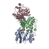
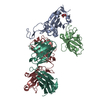
| Title | X-Ray Crystal Structure of the Anthrax Lethal Factor Bound to a Small Molecule Inhibitor, BI-MFM3, 3-{5-[5-(4-Chloro-phenyl)-furan-2-ylmethylene]-4-oxo-2-thioxo-thiazolidin-3-yl}-propionic acid. | ||||||
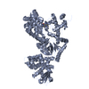
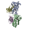
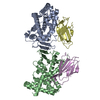
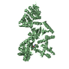
 Components Components | lethal factor | ||||||
 Keywords Keywords | HYDROLASE / Anthrax Toxin Lethal Factor / protein and small molecule inhibitor complex / zinc metalloproteinase | ||||||
| Function / homology |  Function and homology information Function and homology information: / metallopeptidase activity / extracellular region / metal ion binding Similarity search - Function | ||||||
| Biological species |  | ||||||
| Method |  X-RAY DIFFRACTION / MOLECULAR REPLACEMENT / Resolution: 2.67 Å X-RAY DIFFRACTION / MOLECULAR REPLACEMENT / Resolution: 2.67 Å | ||||||
 Authors Authors | Wong, T.Y. / Liddington, R.C. | ||||||
 Citation Citation | Journal: Proc.Natl.Acad.Sci.Usa / Year: 2005 Title: Efficient synthetic inhibitors of anthrax lethal factor. Authors: Forino, M. / Johnson, S. / Wong, T.Y. / Rozanov, D.V. / Savinov, A.Y. / Li, W. / Fattorusso, R. / Becattini, B. / Orry, A.J. / Jung, D. / Abagyan, R.A. / Smith, J.W. / Alibek, K. / ...Authors: Forino, M. / Johnson, S. / Wong, T.Y. / Rozanov, D.V. / Savinov, A.Y. / Li, W. / Fattorusso, R. / Becattini, B. / Orry, A.J. / Jung, D. / Abagyan, R.A. / Smith, J.W. / Alibek, K. / Liddington, R.C. / Strongin, A.Y. / Pellecchia, M. | ||||||
| History |
|
- Structure visualization
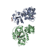
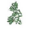
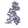
| Structure viewer | Molecule: MolmilJmol/JSmol |
|---|
- Downloads & links
-Download
| PDBx/mmCIF format | 1zxv.cif.gz | 301.9 KB | Display | PDBx/mmCIF format |
|---|---|---|---|---|
| PDB format | pdb1zxv.ent.gz | 245.6 KB | Display | PDB format |
| PDBx/mmJSON format | 1zxv.json.gz | Tree view | PDBx/mmJSON format | |
| Others |  Other downloads Other downloads |
-Validation report
| Summary document | 1zxv_validation.pdf.gz | 968 KB | Display | wwPDB validaton report |
|---|---|---|---|---|
| Full document | 1zxv_full_validation.pdf.gz | 1 MB | Display | |
| Data in XML | 1zxv_validation.xml.gz | 56.7 KB | Display | |
| Data in CIF | 1zxv_validation.cif.gz | 75.8 KB | Display | |
| Arichive directory | https://data.pdbj.org/pub/pdb/validation_reports/zx/1zxvftp://data.pdbj.org/pub/pdb/validation_reports/zx/1zxv | HTTPS FTP |
-Related structure data
| Related structure data |  1j7nS S: Starting model for refinement |
|---|---|
| Similar structure data |

-Links
 PDBj
PDBj


- Assembly
| Deposited unit | 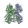
| ||||||||
|---|---|---|---|---|---|---|---|---|---|
| 1 | 
| ||||||||
| 2 | 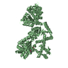
| ||||||||
| Unit cell |
|
-Components
| #1: Protein | Mass: 90356.812 Da / Num. of mol.: 2 / Fragment: Mature peptide Source method: isolated from a genetically manipulated source Source: (gene. exp.) References: GenBank: 143144, UniProt: Q52NH3*PLUS, anthrax lethal factor endopeptidase #2: Chemical |   Mass: 65.409 Da / Num. of mol.: 2 / Source method: obtained synthetically / Formula: Zn Mass: 65.409 Da / Num. of mol.: 2 / Source method: obtained synthetically / Formula: Zn#3: Chemical |   Mass: 393.864 Da / Num. of mol.: 2 / Source method: obtained synthetically / Formula: C17H12ClNO4S2 Mass: 393.864 Da / Num. of mol.: 2 / Source method: obtained synthetically / Formula: C17H12ClNO4S2Details: Chemically synthesised compound based upon initial virtual ligand screening and protein crystal data of initial leads. |
|---|
-Experimental details
-Experiment
| Experiment | Method: X-RAY DIFFRACTION / Number of used crystals: 1 |
|---|
- Sample preparation
| Crystal | Density Matthews: 3.51 Å3/Da / Density % sol: 64.73 % |
|---|---|
| Crystal grow | Temperature: 298 K / Method: vapor diffusion, hanging drop / pH: 8 Details: Ammonium sulfate, Tris-HCl, EDTA., pH 8.0, VAPOR DIFFUSION, HANGING DROP, temperature 298K |
-Data collection
| Diffraction | Mean temperature: 100 K |
|---|---|
| Diffraction source | Source: ROTATING ANODE / Type: RIGAKU FR-E / Wavelength: 1.5418 / Wavelength: 1.5418 Å |
| Detector | Type: RIGAKU RAXIS IV / Detector: IMAGE PLATE / Date: Aug 22, 2004 / Details: Mirrors |
| Radiation | Monochromator: Mirrors / Protocol: SINGLE WAVELENGTH / Monochromatic (M) / Laue (L): M / Scattering type: x-ray |
| Radiation wavelength | Wavelength: 1.5418 Å / Relative weight: 1 |
| Reflection | Resolution: 2.67→49 Å / Num. all: 160988 / Num. obs: 70243 / % possible obs: 98.9 % / Observed criterion σ(I): 1 / Rsym value: 0.074 / Net I/σ(I): 13.29 |
| Reflection shell | Resolution: 2.67→2.77 Å / Mean I/σ(I) obs: 1.76 / Num. unique all: 70243 / Rsym value: 0.5945 / % possible all: 99.6 |
- Processing
| Software |
| |||||||||||||||||||||||||
|---|---|---|---|---|---|---|---|---|---|---|---|---|---|---|---|---|---|---|---|---|---|---|---|---|---|---|
| Refinement | Method to determine structure: MOLECULAR REPLACEMENT Starting model: PDB entry 1J7N Resolution: 2.67→49 Å / Isotropic thermal model: Anisotropic / Cross valid method: THROUGHOUT / σ(F): 0 / σ(I): 1.5
| |||||||||||||||||||||||||
| Refinement step | Cycle: LAST / Resolution: 2.67→49 Å
|
