 Movie
Movie Controller


+ Open data

- Basic information
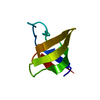
| Entry | Database: PDB / ID: 1zx6 | ||||||
|---|---|---|---|---|---|---|---|
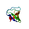
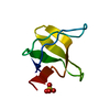
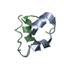
| Title | High-resolution crystal structure of yeast Pin3 SH3 domain | ||||||
 Components Components | Ypr154wp | ||||||
 Keywords Keywords |  PROTEIN BINDING / SH3 domain PROTEIN BINDING / SH3 domain | ||||||
| Function / homology |  Function and homology information Function and homology informationRecycling pathway of L1 / RHOV GTPase cycle / negative regulation of Arp2/3 complex-mediated actin nucleation / actin cortical patch / Antigen processing: Ubiquitination & Proteasome degradation / Neutrophil degranulation / nucleus / cytoplasmSimilarity search - Function | ||||||
| Biological species |  Saccharomyces cerevisiae (brewer's yeast) Saccharomyces cerevisiae (brewer's yeast) | ||||||
| Method | X-RAY DIFFRACTION / SYNCHROTRON / MOLECULAR REPLACEMENT / Resolution: 1.6 Å | ||||||
 Authors Authors | Kursula, P. / Kursula, I. / Lehmann, F. / Zou, P. / Song, Y.H. / Wilmanns, M. | ||||||
 Citation Citation | Journal: To be Published Title: Structural genomics of yeast SH3 domains Authors: Kursula, P. / Kursula, I. / Lehmann, F. / Zou, P. / Song, Y.H. / Wilmanns, M. | ||||||
| History |
|
- Structure visualization
| Structure viewer | Molecule: MolmilJmol/JSmol |
|---|
- Downloads & links
-Download
| PDBx/mmCIF format | 1zx6.cif.gz | 24.5 KB | Display | PDBx/mmCIF format |
|---|---|---|---|---|
| PDB format | pdb1zx6.ent.gz | 15 KB | Display | PDB format |
| PDBx/mmJSON format | 1zx6.json.gz | Tree view | PDBx/mmJSON format | |
| Others |  Other downloads Other downloads |
-Validation report
| Arichive directory | https://data.pdbj.org/pub/pdb/validation_reports/zx/1zx6ftp://data.pdbj.org/pub/pdb/validation_reports/zx/1zx6 | HTTPS FTP |
|---|
-Related structure data
| Related structure data | 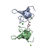 1zukC  1zuuC  2a28C 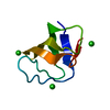 1ootS S: Starting model for refinement C: citing same article ( |
|---|---|
| Similar structure data |


-Links
 PDBj
PDBj


- Assembly
| Deposited unit | 
| ||||||||
|---|---|---|---|---|---|---|---|---|---|
| 1 |
| ||||||||
| Unit cell |
|
-Components
| #1: Protein | Mass: 6516.325 Da / Num. of mol.: 1 / Fragment: SH3 domain Source method: isolated from a genetically manipulated source Source: (gene. exp.) Saccharomyces cerevisiae (brewer's yeast)Plasmid: pDEST-17 / Production host:  Escherichia coli (E. coli) / References: UniProt: Q06449 Escherichia coli (E. coli) / References: UniProt: Q06449 |
|---|---|
| #2: Water | ChemComp-HOH / Water Mass: 18.015 Da / Num. of mol.: 59 / Source method: isolated from a natural source / Formula: H2O Mass: 18.015 Da / Num. of mol.: 59 / Source method: isolated from a natural source / Formula: H2O |
-Experimental details
-Experiment
| Experiment | Method: X-RAY DIFFRACTION / Number of used crystals: 1 |
|---|
- Sample preparation
| Crystal | Density Matthews: 2.2 Å3/Da / Density % sol: 43.5 % |
|---|---|
| Crystal grow | Temperature: 295 K / Method: vapor diffusion, hanging drop / Details: VAPOR DIFFUSION, HANGING DROP, temperature 295K |
-Data collection
| Diffraction | Mean temperature: 100 K |
|---|---|
| Diffraction source | Source: SYNCHROTRON / Site: EMBL/DESY, HAMBURG  / Beamline: X11 / Wavelength: 0.813 Å / Beamline: X11 / Wavelength: 0.813 Å |
| Detector | Type: MARRESEARCH / Detector: CCD / Date: Apr 20, 2005 |
| Radiation | Protocol: SINGLE WAVELENGTH / Monochromatic (M) / Laue (L): M / Scattering type: x-ray |
| Radiation wavelength | Wavelength: 0.813 Å / Relative weight: 1 |
| Reflection | Resolution: 1.6→20 Å / Num. all: 7155 / Num. obs: 7155 / % possible obs: 94.2 % / Observed criterion σ(F): -3 / Observed criterion σ(I): -3 / Redundancy: 2 % / Biso Wilson estimate: 21 Å2 / Rsym value: 0.069 / Net I/σ(I): 7.9 |
| Reflection shell | Resolution: 1.6→1.7 Å / Redundancy: 1.9 % / Mean I/σ(I) obs: 2.6 / Num. unique all: 1187 / Rsym value: 0.312 / % possible all: 96.8 |
- Processing
| Software |
| |||||||||||||||||||||||||||||||||||||||||||||||||||||||||||||||||||||||||||||||||||||||||||||||||||||||||||||||||||||||||||||
|---|---|---|---|---|---|---|---|---|---|---|---|---|---|---|---|---|---|---|---|---|---|---|---|---|---|---|---|---|---|---|---|---|---|---|---|---|---|---|---|---|---|---|---|---|---|---|---|---|---|---|---|---|---|---|---|---|---|---|---|---|---|---|---|---|---|---|---|---|---|---|---|---|---|---|---|---|---|---|---|---|---|---|---|---|---|---|---|---|---|---|---|---|---|---|---|---|---|---|---|---|---|---|---|---|---|---|---|---|---|---|---|---|---|---|---|---|---|---|---|---|---|---|---|---|---|---|
| Refinement | Method to determine structure: MOLECULAR REPLACEMENT Starting model: PDB Entry: 1oot Resolution: 1.6→20 Å / Cor.coef. Fo:Fc: 0.961 / Cor.coef. Fo:Fc free: 0.938 / SU B: 4.335 / SU ML: 0.073 / TLS residual ADP flag: LIKELY RESIDUAL / Isotropic thermal model: TLS REFINEMENT / Cross valid method: THROUGHOUT / σ(F): -3 / ESU R: 0.098 / ESU R Free: 0.1 / Stereochemistry target values: Engh & Huber / Details: HYDROGENS HAVE BEEN ADDED IN THE RIDING POSITIONS
| |||||||||||||||||||||||||||||||||||||||||||||||||||||||||||||||||||||||||||||||||||||||||||||||||||||||||||||||||||||||||||||
| Solvent computation | Ion probe radii: 0.8 Å / Shrinkage radii: 0.8 Å / VDW probe radii: 1.2 Å / Solvent model: BABINET MODEL WITH MASK | |||||||||||||||||||||||||||||||||||||||||||||||||||||||||||||||||||||||||||||||||||||||||||||||||||||||||||||||||||||||||||||
| Displacement parameters | Biso mean: 21.942 Å2
| |||||||||||||||||||||||||||||||||||||||||||||||||||||||||||||||||||||||||||||||||||||||||||||||||||||||||||||||||||||||||||||
| Refinement step | Cycle: LAST / Resolution: 1.6→20 Å
| |||||||||||||||||||||||||||||||||||||||||||||||||||||||||||||||||||||||||||||||||||||||||||||||||||||||||||||||||||||||||||||
| Refine LS restraints |
| |||||||||||||||||||||||||||||||||||||||||||||||||||||||||||||||||||||||||||||||||||||||||||||||||||||||||||||||||||||||||||||
| LS refinement shell | Resolution: 1.6→1.641 Å / Total num. of bins used: 20
| |||||||||||||||||||||||||||||||||||||||||||||||||||||||||||||||||||||||||||||||||||||||||||||||||||||||||||||||||||||||||||||
| Refinement TLS params. | Method: refined / Origin x: 12.1845 Å / Origin y: 27.5746 Å / Origin z: 3.0117 Å
|
