 Movie
Movie Controller

[English] 日本語
 Yorodumi
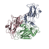
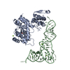
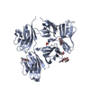
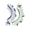
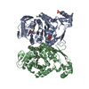
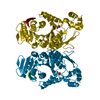
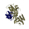
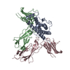
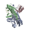
- PDB-1zs4: Structure of bacteriophage lambda cII protein in complex with DNA -
+ Open data

- Basic information
| Entry | Database: PDB / ID: 1zs4 | ||||||
|---|---|---|---|---|---|---|---|
| Title | Structure of bacteriophage lambda cII protein in complex with DNA | ||||||
 Components Components |
| ||||||
 Keywords Keywords | transcription/DNA / HELIX-TURN-HELIX / TRANSCRIPTION ACTIVATOR / transcription-DNA COMPLEX | ||||||
| Function / homology |  Function and homology information Function and homology information | ||||||
| Biological species |  Enterobacteria phage lambda (virus) Enterobacteria phage lambda (virus) | ||||||
| Method |  X-RAY DIFFRACTION / SYNCHROTRON / MAD / Resolution: 1.7 Å X-RAY DIFFRACTION / SYNCHROTRON / MAD / Resolution: 1.7 Å | ||||||
 Authors Authors | Jain, D. / Kim, Y. / Maxwell, K.L. / Beasley, S. / Gussin, G.N. / Edwards, A.M. / Darst, S.A. | ||||||
 Citation Citation | Journal: Mol.Cell / Year: 2005 Title: Crystal Structure of Bacteriophage lambdacII and Its DNA Complex. Authors: Jain, D. / Kim, Y. / Maxwell, K.L. / Beasley, S. / Zhang, R. / Gussin, G.N. / Edwards, A.M. / Darst, S.A. | ||||||
| History |
|
- Structure visualization
| Structure viewer | Molecule: MolmilJmol/JSmol |
|---|
- Downloads & links
-Download
| PDBx/mmCIF format | 1zs4.cif.gz | 110.6 KB | Display | PDBx/mmCIF format |
|---|---|---|---|---|
| PDB format | pdb1zs4.ent.gz | 82.7 KB | Display | PDB format |
| PDBx/mmJSON format | 1zs4.json.gz | Tree view | PDBx/mmJSON format | |
| Others |  Other downloads Other downloads |
-Validation report
| Summary document | 1zs4_validation.pdf.gz | 467.1 KB | Display | wwPDB validaton report |
|---|---|---|---|---|
| Full document | 1zs4_full_validation.pdf.gz | 476.2 KB | Display | |
| Data in XML | 1zs4_validation.xml.gz | 20.2 KB | Display | |
| Data in CIF | 1zs4_validation.cif.gz | 30.7 KB | Display | |
| Arichive directory | https://data.pdbj.org/pub/pdb/validation_reports/zs/1zs4ftp://data.pdbj.org/pub/pdb/validation_reports/zs/1zs4 | HTTPS FTP |
-Related structure data


-Links
 PDBj
PDBj


- Assembly
| Deposited unit | 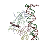
| ||||||||
|---|---|---|---|---|---|---|---|---|---|
| 1 |
| ||||||||
| Unit cell |
|
-Components
| #1: DNA chain | Mass: 8264.316 Da / Num. of mol.: 1 / Source method: obtained synthetically / Details: SYNTHETIC | ||
|---|---|---|---|
| #2: DNA chain | Mass: 8318.400 Da / Num. of mol.: 1 / Source method: obtained synthetically / Details: SYNTHETIC | ||
| #3: Protein | Mass: 9321.896 Da / Num. of mol.: 4 Source method: isolated from a genetically manipulated source Source: (gene. exp.) Enterobacteria phage lambda (virus) / Genus: Lambda-like viruses / Gene: CII / Plasmid: pET15b / Species (production host): Escherichia coli / Production host:  #4: Water | ChemComp-HOH / |  Mass: 18.015 Da / Num. of mol.: 417 / Source method: isolated from a natural source / Formula: H2O Mass: 18.015 Da / Num. of mol.: 417 / Source method: isolated from a natural source / Formula: H2O |
-Experimental details
-Experiment
| Experiment | Method: X-RAY DIFFRACTION / Number of used crystals: 1 |
|---|
- Sample preparation
| Crystal | Density Matthews: 2.3 Å3/Da / Density % sol: 46.61 % | ||||||||||||||||
|---|---|---|---|---|---|---|---|---|---|---|---|---|---|---|---|---|---|
| Crystal grow | Temperature: 295 K / Method: vapor diffusion / pH: 4.8 Details: PEG 550 MME, SODIUM ACETATE, pH 4.8, VAPOR DIFFUSION, temperature 295K | ||||||||||||||||
| Components of the solutions |
|
-Data collection
| Diffraction source | Source: SYNCHROTRON / Site: NSLS  / Beamline: X9A / Wavelength: 0.98396, 0.97903, 0.97922, 0.96384 / Beamline: X9A / Wavelength: 0.98396, 0.97903, 0.97922, 0.96384 | |||||||||||||||
|---|---|---|---|---|---|---|---|---|---|---|---|---|---|---|---|---|
| Detector | Type: MARRESEARCH / Detector: CCD / Date: Aug 24, 2004 | |||||||||||||||
| Radiation | Monochromator: Si 111 / Protocol: MAD / Monochromatic (M) / Laue (L): M / Scattering type: x-ray | |||||||||||||||
| Radiation wavelength |
| |||||||||||||||
| Reflection | Resolution: 1.7→50 Å / Num. obs: 54170 / % possible obs: 97.3 % / Rsym value: 0.067 / Net I/σ(I): 8.7 |
- Processing
| Software |
| ||||||||||||||||||||
|---|---|---|---|---|---|---|---|---|---|---|---|---|---|---|---|---|---|---|---|---|---|
| Refinement | Method to determine structure: MAD / Resolution: 1.7→50 Å / σ(F): 0
| ||||||||||||||||||||
| Refinement step | Cycle: LAST / Resolution: 1.7→50 Å
|
