| 登録情報 | データベース: PDB / ID: 7dsb
|
|---|
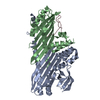
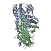
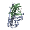
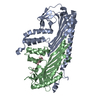
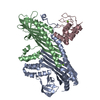
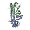
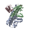
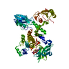
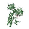
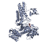
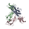
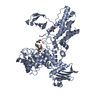
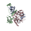
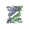
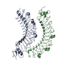
| タイトル | Crystal structure of actin capping protein in complex with V-1 and twinfilin C-terminal tail |
|---|
 要素 要素 | - F-actin-capping protein subunit alpha-1
- F-actin-capping protein subunit beta isoforms 1
- Myotrophin
 MTPN MTPN - Twinfilin-1
|
|---|
 キーワード キーワード | CYTOSOLIC PROTEIN (細胞質基質) / actin dynamics / actin capping protein / twinfilin / CARMIL / V-1 |
|---|
| 機能・相同性 |  機能・相同性情報 機能・相同性情報
positive regulation of macromolecule biosynthetic process / regulation of barbed-end actin filament capping / regulation of actin phosphorylation / Advanced glycosylation endproduct receptor signaling / RHOD GTPase cycle / RHOF GTPase cycle / HSP90 chaperone cycle for steroid hormone receptors (SHR) in the presence of ligand / COPI-independent Golgi-to-ER retrograde traffic / RHOBTB2 GTPase cycle / COPI-mediated anterograde transport ...positive regulation of macromolecule biosynthetic process / regulation of barbed-end actin filament capping / regulation of actin phosphorylation / Advanced glycosylation endproduct receptor signaling / RHOD GTPase cycle / RHOF GTPase cycle / HSP90 chaperone cycle for steroid hormone receptors (SHR) in the presence of ligand / COPI-independent Golgi-to-ER retrograde traffic / RHOBTB2 GTPase cycle / COPI-mediated anterograde transport / Factors involved in megakaryocyte development and platelet production / cerebellar granule cell differentiation / regulation of striated muscle tissue development / WASH complex / sperm connecting piece / F-actin capping protein complex / negative regulation of filopodium assembly / sequestering of actin monomers / skeletal muscle tissue regeneration / negative regulation of actin filament polymerization / catecholamine metabolic process / cell junction assembly / barbed-end actin filament capping / actin polymerization or depolymerization / actin filament depolymerization / regulation of cell morphogenesis / regulation of lamellipodium assembly / lamellipodium assembly / regulation of cell size / myofibril / cortical cytoskeleton / positive regulation of cardiac muscle hypertrophy / 刷子縁 / actin monomer binding / cytoskeleton organization / striated muscle cell differentiation / phosphatidylinositol-4,5-bisphosphate binding / hippocampal mossy fiber to CA3 synapse / positive regulation of protein metabolic process / filopodium / マイクロフィラメント / Schaffer collateral - CA1 synapse / cell morphogenesis / neuron differentiation / Z disc / positive regulation of neuron projection development / cellular response to mechanical stimulus / actin filament binding / cell-cell junction / マイクロフィラメント / regulation of translation / lamellipodium / positive regulation of NF-kappaB transcription factor activity / actin binding / actin cytoskeleton organization / positive regulation of cell growth / protein tyrosine kinase activity / sequence-specific DNA binding / postsynaptic density / 神経繊維 / protein-containing complex binding / perinuclear region of cytoplasm / ATP binding / 生体膜 / 細胞核 / 細胞膜 / 細胞質基質 / 細胞質類似検索 - 分子機能 Twinfilin / F-actin-capping protein subunit beta / F-actin capping protein, beta subunit, conserved site / F-actin-capping protein subunit beta, N-terminal domain / F-actin capping protein, beta subunit / F-actin capping protein beta subunit signature. / F-actin capping protein, alpha subunit, conserved site / F-actin capping protein alpha subunit signature 1. / F-actin capping protein alpha subunit signature 2. / F-actin-capping protein subunit alpha ...Twinfilin / F-actin-capping protein subunit beta / F-actin capping protein, beta subunit, conserved site / F-actin-capping protein subunit beta, N-terminal domain / F-actin capping protein, beta subunit / F-actin capping protein beta subunit signature. / F-actin capping protein, alpha subunit, conserved site / F-actin capping protein alpha subunit signature 1. / F-actin capping protein alpha subunit signature 2. / F-actin-capping protein subunit alpha / F-actin-capping protein subunit alpha/beta / F-actin-capping protein subunit alpha/beta, domain 2 / F-actin capping protein, alpha subunit, domain 1 / F-actin capping protein alpha subunit / Actin-depolymerising factor homology domain / Cofilin/tropomyosin-type actin-binding protein / ADF-H domain profile. / Actin depolymerisation factor/cofilin -like domains / ADF-H/Gelsolin-like domain superfamily / Ankyrin repeats (3 copies) / Ankyrin repeat profile. / Ankyrin repeat region circular profile. / ankyrin repeats / Ankyrin repeat / Ankyrin repeat-containing domain superfamily類似検索 - ドメイン・相同性 F-actin-capping protein subunit alpha-1 / F-actin-capping protein subunit beta isoforms 1 and 2 / Myotrophin / Twinfilin-1類似検索 - 構成要素 |
|---|
| 生物種 |   Gallus gallus (ニワトリ) Gallus gallus (ニワトリ)
 Homo sapiens (ヒト) Homo sapiens (ヒト)
  Mus musculus (ハツカネズミ) Mus musculus (ハツカネズミ) |
|---|
| 手法 | X線回折 / シンクロトロン / 分子置換 / 解像度: 2.44 Å |
|---|
 データ登録者 データ登録者 | Takeda, S. |
|---|
| 資金援助 |  日本, 3件 日本, 3件 | 組織 | 認可番号 | 国 |
|---|
| Japan Society for the Promotion of Science (JSPS) | 16K17708 | 日本 | | Japan Society for the Promotion of Science (JSPS) | 17K07373 | 日本 | | Japan Society for the Promotion of Science (JSPS) | 20K06522 | 日本 |
|
|---|
 引用 引用 | ジャーナル: J.Mol.Biol. / 年: 2021
タイトル: Structural Insights into the Regulation of Actin Capping Protein by Twinfilin C-terminal Tail.
著者: Takeda, S. / Koike, R. / Fujiwara, I. / Narita, A. / Miyata, M. / Ota, M. / Maeda, Y. |
|---|
| 履歴 | | 登録 | 2020年12月30日 | 登録サイト: PDBJ / 処理サイト: PDBJ |
|---|
| 改定 1.0 | 2021年3月3日 | Provider: repository / タイプ: Initial release |
|---|
| 改定 1.1 | 2021年3月17日 | Group: Database references / カテゴリ: citation / citation_author
Item: _citation.page_first / _citation.page_last ..._citation.page_first / _citation.page_last / _citation.pdbx_database_id_PubMed / _citation.title / _citation_author.identifier_ORCID |
|---|
| 改定 1.2 | 2021年4月7日 | Group: Database references / カテゴリ: citation / Item: _citation.journal_volume / _citation.title |
|---|
| 改定 1.3 | 2023年11月29日 | Group: Data collection / Database references / Refinement description
カテゴリ: chem_comp_atom / chem_comp_bond ...chem_comp_atom / chem_comp_bond / database_2 / pdbx_initial_refinement_model
Item: _database_2.pdbx_DOI / _database_2.pdbx_database_accession |
|---|
|
|---|
 ムービー
ムービー コントローラー


 データを開く
 基本情報
基本情報 構造の表示
構造の表示 ダウンロードとリンク
 その他のダウンロード
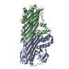

 PDBj
PDBj
 集合体
集合体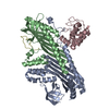

 分子量: 18.015 Da / 分子数: 94 / 由来タイプ: 天然 / 式: H2O
 試料調製
試料調製 解析
解析