 Movie
Movie Controller

[English] 日本語
 Yorodumi
- PDB-5d4k: Crystal structure of the human polymeric Ig receptor (pIgR) ectodomain -
+ Open data

- Basic information
| Entry | Database: PDB / ID: 5d4k | |||||||||
|---|---|---|---|---|---|---|---|---|---|---|
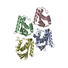
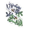
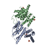
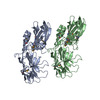
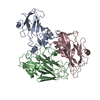
| Title | Crystal structure of the human polymeric Ig receptor (pIgR) ectodomain | |||||||||
 Components Components | Polymeric immunoglobulin receptor | |||||||||
 Keywords Keywords | IMMUNE SYSTEM / Ig Super Family (IgSF) / polymeric Ig-binding protein / mucosal immunity / Secretory Component | |||||||||
| Function / homology |  Function and homology informationpolymeric immunoglobulin receptor activity / immunoglobulin transcytosis in epithelial cells mediated by polymeric immunoglobulin receptor / polymeric immunoglobulin binding / secretory IgA immunoglobulin complex / Fc receptor signaling pathway / detection of chemical stimulus involved in sensory perception of bitter taste / azurophil granule membrane / receptor clustering / epidermal growth factor receptor signaling pathway / receptor complex ...polymeric immunoglobulin receptor activity / immunoglobulin transcytosis in epithelial cells mediated by polymeric immunoglobulin receptor / polymeric immunoglobulin binding / secretory IgA immunoglobulin complex / Fc receptor signaling pathway / detection of chemical stimulus involved in sensory perception of bitter taste / azurophil granule membrane / receptor clustering / epidermal growth factor receptor signaling pathway / receptor complex / Neutrophil degranulation / extracellular space / extracellular exosome / plasma membrane Function and homology informationpolymeric immunoglobulin receptor activity / immunoglobulin transcytosis in epithelial cells mediated by polymeric immunoglobulin receptor / polymeric immunoglobulin binding / secretory IgA immunoglobulin complex / Fc receptor signaling pathway / detection of chemical stimulus involved in sensory perception of bitter taste / azurophil granule membrane / receptor clustering / epidermal growth factor receptor signaling pathway / receptor complex ...polymeric immunoglobulin receptor activity / immunoglobulin transcytosis in epithelial cells mediated by polymeric immunoglobulin receptor / polymeric immunoglobulin binding / secretory IgA immunoglobulin complex / Fc receptor signaling pathway / detection of chemical stimulus involved in sensory perception of bitter taste / azurophil granule membrane / receptor clustering / epidermal growth factor receptor signaling pathway / receptor complex / Neutrophil degranulation / extracellular space / extracellular exosome / plasma membraneSimilarity search - Function | |||||||||
| Biological species |  Homo sapiens (human) Homo sapiens (human) | |||||||||
| Method | X-RAY DIFFRACTION / SYNCHROTRON / MOLECULAR REPLACEMENT / Resolution: 2.599 Å | |||||||||
 Authors Authors | Stadtmueller, B.M. / Bjorkman, P.J. | |||||||||
| Funding support |  United States, 2items United States, 2items
| |||||||||
 Citation Citation | Journal: Elife / Year: 2016 Title: The structure and dynamics of secretory component and its interactions with polymeric immunoglobulins. Authors: Stadtmueller, B.M. / Huey-Tubman, K.E. / Lopez, C.J. / Yang, Z. / Hubbell, W.L. / Bjorkman, P.J. | |||||||||
| History |
|
- Structure visualization
| Structure viewer | Molecule: MolmilJmol/JSmol |
|---|
- Downloads & links
-Download
| PDBx/mmCIF format | 5d4k.cif.gz | 398.6 KB | Display | PDBx/mmCIF format |
|---|---|---|---|---|
| PDB format | pdb5d4k.ent.gz | 331.9 KB | Display | PDB format |
| PDBx/mmJSON format | 5d4k.json.gz | Tree view | PDBx/mmJSON format | |
| Others |  Other downloads Other downloads |
-Validation report
| Arichive directory | https://data.pdbj.org/pub/pdb/validation_reports/d4/5d4kftp://data.pdbj.org/pub/pdb/validation_reports/d4/5d4k | HTTPS FTP |
|---|
-Related structure data
| Related structure data |  5f1sC  1xedS C: citing same article ( S: Starting model for refinement |
|---|---|
| Similar structure data |


-Links
 PDBj
PDBj- Assembly
| Deposited unit | 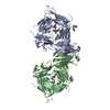
| |||||||||||||||||||||
|---|---|---|---|---|---|---|---|---|---|---|---|---|---|---|---|---|---|---|---|---|---|---|
| 1 | 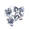
| |||||||||||||||||||||
| 2 | 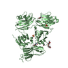
| |||||||||||||||||||||
| Unit cell |
| |||||||||||||||||||||
| Noncrystallographic symmetry (NCS) | NCS domain:
NCS domain segments: Component-ID: 1 / Ens-ID: 1 / Beg auth comp-ID: SER / Beg label comp-ID: SER / End auth comp-ID: SER / End label comp-ID: SER / Auth seq-ID: 2 - 549 / Label seq-ID: 2 - 549
|
-Components
| #1: Protein | / Poly-Ig receptor / Hepatocellular carcinoma-associated protein TB6 Mass: 61483.004 Da / Num. of mol.: 2 / Fragment: ectodomain (UNP residues 19-565) Source method: isolated from a genetically manipulated source Source: (gene. exp.) Homo sapiens (human) / Gene: PIGR / Plasmid: pTT5 / Cell line (production host): HEK 293-6E / Production host: Homo sapiens (human) / References: UniProt: P01833#2: Polysaccharide | / Mass: 367.349 Da / Num. of mol.: 2Source method: isolated from a genetically manipulated source #3: Sugar | ChemComp-NAG / N-Acetylglucosamine  Type: D-saccharide, beta linking / Mass: 221.208 Da / Num. of mol.: 6 Type: D-saccharide, beta linking / Mass: 221.208 Da / Num. of mol.: 6Source method: isolated from a genetically manipulated source Formula: C8H15NO6 #4: Chemical | ChemComp-SO4 / Sulfate  Mass: 96.063 Da / Num. of mol.: 4 / Source method: obtained synthetically / Formula: SO4 Mass: 96.063 Da / Num. of mol.: 4 / Source method: obtained synthetically / Formula: SO4#5: Water | ChemComp-HOH / | Water Mass: 18.015 Da / Num. of mol.: 58 / Source method: isolated from a natural source / Formula: H2O Mass: 18.015 Da / Num. of mol.: 58 / Source method: isolated from a natural source / Formula: H2O |
|---|
-Experimental details
-Experiment
| Experiment | Method: X-RAY DIFFRACTION / Number of used crystals: 1 |
|---|
- Sample preparation
| Crystal | Density Matthews: 3.45 Å3/Da / Density % sol: 64.38 % |
|---|---|
| Crystal grow | Temperature: 295 K / Method: vapor diffusion, sitting drop / pH: 5.5 Details: 0.2 M ammonium sulfate, 0.1M BisTris pH 5.5, 31% Polyethylene glycol 3350 |
-Data collection
| Diffraction | Mean temperature: 100 K | ||||||||||||||||||||||||||||||||||||||||||||||||||||||||||||||||||||||||||||||||||||||||||||||||||||||||||||||
|---|---|---|---|---|---|---|---|---|---|---|---|---|---|---|---|---|---|---|---|---|---|---|---|---|---|---|---|---|---|---|---|---|---|---|---|---|---|---|---|---|---|---|---|---|---|---|---|---|---|---|---|---|---|---|---|---|---|---|---|---|---|---|---|---|---|---|---|---|---|---|---|---|---|---|---|---|---|---|---|---|---|---|---|---|---|---|---|---|---|---|---|---|---|---|---|---|---|---|---|---|---|---|---|---|---|---|---|---|---|---|---|
| Diffraction source | Source: SYNCHROTRON / Site: SSRL / Beamline: BL12-2 / Wavelength: 0.9537 Å | ||||||||||||||||||||||||||||||||||||||||||||||||||||||||||||||||||||||||||||||||||||||||||||||||||||||||||||||
| Detector | Type: DECTRIS PILATUS 6M / Detector: PIXEL / Date: May 10, 2012 | ||||||||||||||||||||||||||||||||||||||||||||||||||||||||||||||||||||||||||||||||||||||||||||||||||||||||||||||
| Radiation | Monochromator: Liquid nitrogen-cooled double crystal Si(111) Protocol: SINGLE WAVELENGTH / Monochromatic (M) / Laue (L): M / Scattering type: x-ray | ||||||||||||||||||||||||||||||||||||||||||||||||||||||||||||||||||||||||||||||||||||||||||||||||||||||||||||||
| Radiation wavelength | Wavelength: 0.9537 Å / Relative weight: 1 | ||||||||||||||||||||||||||||||||||||||||||||||||||||||||||||||||||||||||||||||||||||||||||||||||||||||||||||||
| Reflection | Resolution: 2.599→39.623 Å / Num. all: 49242 / Num. obs: 49242 / % possible obs: 96.5 % / Redundancy: 3 % / Biso Wilson estimate: 51.53 Å2 / Rpim(I) all: 0.039 / Rrim(I) all: 0.07 / Rsym value: 0.058 / Net I/av σ(I): 11.696 / Net I/σ(I): 13.7 / Num. measured all: 145998 | ||||||||||||||||||||||||||||||||||||||||||||||||||||||||||||||||||||||||||||||||||||||||||||||||||||||||||||||
| Reflection shell | Diffraction-ID: 1 / Rejects: 0
|
- Processing
| Software |
| |||||||||||||||||||||||||||||||||||||||||||||||||||||||||||||||||||||||||||||||||||||||||||||||||||||||||
|---|---|---|---|---|---|---|---|---|---|---|---|---|---|---|---|---|---|---|---|---|---|---|---|---|---|---|---|---|---|---|---|---|---|---|---|---|---|---|---|---|---|---|---|---|---|---|---|---|---|---|---|---|---|---|---|---|---|---|---|---|---|---|---|---|---|---|---|---|---|---|---|---|---|---|---|---|---|---|---|---|---|---|---|---|---|---|---|---|---|---|---|---|---|---|---|---|---|---|---|---|---|---|---|---|---|---|
| Refinement | Method to determine structure: MOLECULAR REPLACEMENT Starting model: 1XED Resolution: 2.599→36.984 Å / SU ML: 0.37 / Cross valid method: FREE R-VALUE / σ(F): 0.02 / Phase error: 28.17 / Stereochemistry target values: ML
| |||||||||||||||||||||||||||||||||||||||||||||||||||||||||||||||||||||||||||||||||||||||||||||||||||||||||
| Solvent computation | Shrinkage radii: 0.9 Å / VDW probe radii: 1.11 Å / Solvent model: FLAT BULK SOLVENT MODEL | |||||||||||||||||||||||||||||||||||||||||||||||||||||||||||||||||||||||||||||||||||||||||||||||||||||||||
| Displacement parameters | Biso max: 271.63 Å2 / Biso mean: 69.2157 Å2 / Biso min: 22.82 Å2 | |||||||||||||||||||||||||||||||||||||||||||||||||||||||||||||||||||||||||||||||||||||||||||||||||||||||||
| Refinement step | Cycle: final / Resolution: 2.599→36.984 Å
| |||||||||||||||||||||||||||||||||||||||||||||||||||||||||||||||||||||||||||||||||||||||||||||||||||||||||
| Refine LS restraints |
| |||||||||||||||||||||||||||||||||||||||||||||||||||||||||||||||||||||||||||||||||||||||||||||||||||||||||
| Refine LS restraints NCS |
| |||||||||||||||||||||||||||||||||||||||||||||||||||||||||||||||||||||||||||||||||||||||||||||||||||||||||
| LS refinement shell | Refine-ID: X-RAY DIFFRACTION / Total num. of bins used: 14
|
