 Movie
Movie Controller

[English] 日本語
 Yorodumi
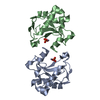
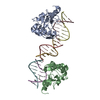
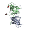
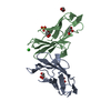
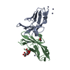
- PDB-1znv: How a His-metal finger endonuclease ColE7 binds and cleaves DNA w... -
+ Open data

- Basic information
| Entry | Database: PDB / ID: 1znv | ||||||
|---|---|---|---|---|---|---|---|
| Title | How a His-metal finger endonuclease ColE7 binds and cleaves DNA with a transition metal ion cofactor | ||||||
 Components Components |
| ||||||
 Keywords Keywords | Hydrolase/PROTEIN BINDING / H-N-H motif / Ni-binding / protein-protein complex /  endonuclease / Hydrolase-PROTEIN BINDING COMPLEX endonuclease / Hydrolase-PROTEIN BINDING COMPLEX | ||||||
| Function / homology |  Function and homology informationextrachromosomal circular DNA / bacteriocin immunity / toxic substance binding / endonuclease activity / killing of cells of another organism / Hydrolases; Acting on ester bonds / defense response to bacterium / metal ion binding Function and homology informationextrachromosomal circular DNA / bacteriocin immunity / toxic substance binding / endonuclease activity / killing of cells of another organism / Hydrolases; Acting on ester bonds / defense response to bacterium / metal ion bindingSimilarity search - Function | ||||||
| Biological species |  Escherichia coli str. K12 substr. (bacteria) Escherichia coli str. K12 substr. (bacteria) | ||||||
| Method | X-RAY DIFFRACTION / SYNCHROTRON / Isomorphous to 1MZ8 / Resolution: 2 Å | ||||||
 Authors Authors | Doudeva, L.G. / Huang, H. / Hsia, K.C. / Shi, Z. / Li, C.L. / Shen, Y. / Yuan, H.S. | ||||||
 Citation Citation | Journal: Protein Sci. / Year: 2006 Title: Crystal structural analysis and metal-dependent stability and activity studies of the ColE7 endonuclease domain in complex with DNA/Zn2+ or inhibitor/Ni2+ Authors: Doudeva, L.G. / Huang, H. / Hsia, K.C. / Shi, Z. / Li, C.L. / Shen, Y. / Cheng, Y.S. / Yuan, H.S. | ||||||
| History |
|
- Structure visualization
| Structure viewer | Molecule: MolmilJmol/JSmol |
|---|
- Downloads & links
-Download
| PDBx/mmCIF format | 1znv.cif.gz | 107 KB | Display | PDBx/mmCIF format |
|---|---|---|---|---|
| PDB format | pdb1znv.ent.gz | 81 KB | Display | PDB format |
| PDBx/mmJSON format | 1znv.json.gz | Tree view | PDBx/mmJSON format | |
| Others |  Other downloads Other downloads |
-Validation report
| Arichive directory | https://data.pdbj.org/pub/pdb/validation_reports/zn/1znvftp://data.pdbj.org/pub/pdb/validation_reports/zn/1znv | HTTPS FTP |
|---|
-Related structure data
| Related structure data | 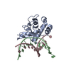 1znsC 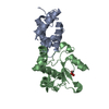 1mz8S S: Starting model for refinement C: citing same article ( |
|---|---|
| Similar structure data |


-Links
 PDBj
PDBj
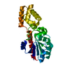
- Assembly
| Deposited unit | 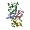
| ||||||||
|---|---|---|---|---|---|---|---|---|---|
| 1 | 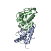
| ||||||||
| 2 | 
| ||||||||
| Unit cell |
|
-Components
| #1: Protein | Mass: 10735.845 Da / Num. of mol.: 2 Source method: isolated from a genetically manipulated source Source: (gene. exp.) Escherichia coli str. K12 substr. (bacteria)Species: Escherichia coli / Strain: W3110 / Gene: cei7 / Plasmid: pQE70 / Production host: Escherichia coli (E. coli) / Strain (production host): M15 / References: UniProt: Q03708#2: Protein | Mass: 15400.457 Da / Num. of mol.: 2 / Fragment: Nuclease domain / Mutation: H545E Source method: isolated from a genetically manipulated source Source: (gene. exp.) Escherichia coli str. K12 substr. (bacteria)Species: Escherichia coli / Strain: W3110 / Gene: cei7 / Plasmid: pQE70 / Production host: Escherichia coli (E. coli) / Strain (production host): M15References: UniProt: Q47112, Hydrolases; Acting on ester bonds#3: Chemical | Nickel  Mass: 58.693 Da / Num. of mol.: 2 / Source method: obtained synthetically / Formula: Ni Mass: 58.693 Da / Num. of mol.: 2 / Source method: obtained synthetically / Formula: Ni#4: Chemical | Phosphate  Mass: 94.971 Da / Num. of mol.: 2 / Source method: obtained synthetically / Formula: PO4 Mass: 94.971 Da / Num. of mol.: 2 / Source method: obtained synthetically / Formula: PO4#5: Water | ChemComp-HOH / | Water Mass: 18.015 Da / Num. of mol.: 423 / Source method: isolated from a natural source / Formula: H2O Mass: 18.015 Da / Num. of mol.: 423 / Source method: isolated from a natural source / Formula: H2O |
|---|
-Experimental details
-Experiment
| Experiment | Method: X-RAY DIFFRACTION / Number of used crystals: 1 |
|---|
- Sample preparation
| Crystal | Density Matthews: 2.8 Å3/Da / Density % sol: 56.02 % |
|---|---|
| Crystal grow | Temperature: 298 K / Method: vapor diffusion, hanging drop / pH: 5.7 Details: 0.3M phosphate buffer, 50mM NaCl, 20% PEG550 MME, 10% glycerol, pH 5.7, VAPOR DIFFUSION, HANGING DROP, temperature 298K |
-Data collection
| Diffraction | Mean temperature: 120 K |
|---|---|
| Diffraction source | Source: SYNCHROTRON / Site: SPring-8  / Beamline: BL12B2 / Wavelength: 1 Å / Beamline: BL12B2 / Wavelength: 1 Å |
| Detector | Type: ADSC QUANTUM 4 / Detector: CCD / Date: Dec 15, 2004 |
| Radiation | Monochromator: double crystal monochromator (DCM) / Protocol: SINGLE WAVELENGTH / Monochromatic (M) / Laue (L): M / Scattering type: x-ray |
| Radiation wavelength | Wavelength: 1 Å / Relative weight: 1 |
| Reflection | Resolution: 2→40 Å / Num. all: 224307 / Num. obs: 223845 / % possible obs: 99.8 % / Redundancy: 5.8 % / Biso Wilson estimate: 16.3 Å2 / Rsym value: 0.082 / Net I/σ(I): 25.34 |
| Reflection shell | Resolution: 2→2.07 Å / Mean I/σ(I) obs: 5.73 / Num. unique all: 4763 / Rsym value: 0.381 |
- Processing
| Software |
| ||||||||||||||||||||||||||||||||||||||||||||||||||||||||||||||||||||||||||||||||
|---|---|---|---|---|---|---|---|---|---|---|---|---|---|---|---|---|---|---|---|---|---|---|---|---|---|---|---|---|---|---|---|---|---|---|---|---|---|---|---|---|---|---|---|---|---|---|---|---|---|---|---|---|---|---|---|---|---|---|---|---|---|---|---|---|---|---|---|---|---|---|---|---|---|---|---|---|---|---|---|---|---|
| Refinement | Method to determine structure: Isomorphous to 1MZ8 Starting model: PDB ENTRY 1MZ8 Resolution: 2→27.65 Å / Rfactor Rfree error: 0.004 / Data cutoff high absF: 412157.16 / Data cutoff low absF: 0 / Isotropic thermal model: RESTRAINED / Cross valid method: THROUGHOUT / σ(F): 0 / Stereochemistry target values: Engh & Huber
| ||||||||||||||||||||||||||||||||||||||||||||||||||||||||||||||||||||||||||||||||
| Solvent computation | Solvent model: FLAT MODEL / Bsol: 58.7927 Å2 / ksol: 0.374744 e/Å3 | ||||||||||||||||||||||||||||||||||||||||||||||||||||||||||||||||||||||||||||||||
| Displacement parameters | Biso mean: 32.8 Å2
| ||||||||||||||||||||||||||||||||||||||||||||||||||||||||||||||||||||||||||||||||
| Refine analyze |
| ||||||||||||||||||||||||||||||||||||||||||||||||||||||||||||||||||||||||||||||||
| Refinement step | Cycle: LAST / Resolution: 2→27.65 Å
| ||||||||||||||||||||||||||||||||||||||||||||||||||||||||||||||||||||||||||||||||
| Refine LS restraints |
| ||||||||||||||||||||||||||||||||||||||||||||||||||||||||||||||||||||||||||||||||
| LS refinement shell | Resolution: 2→2.09 Å / Rfactor Rfree error: 0.012 / Total num. of bins used: 6
| ||||||||||||||||||||||||||||||||||||||||||||||||||||||||||||||||||||||||||||||||
| Xplor file |
|
