 Movie
Movie Controller


+ Open data

- Basic information
| Entry | Database: PDB / ID: 1m08 | ||||||
|---|---|---|---|---|---|---|---|
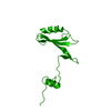
| Title | Crystal structure of the unbound nuclease domain of ColE7 | ||||||
 Components Components | Colicin E7 | ||||||
 Keywords Keywords |  HYDROLASE / HNH motif / endonuclease / colicin / Zn-binding protein HYDROLASE / HNH motif / endonuclease / colicin / Zn-binding protein | ||||||
| Function / homology |  Function and homology informationextrachromosomal circular DNA / endonuclease activity / killing of cells of another organism / Hydrolases; Acting on ester bonds / defense response to bacterium / metal ion binding Function and homology informationextrachromosomal circular DNA / endonuclease activity / killing of cells of another organism / Hydrolases; Acting on ester bonds / defense response to bacterium / metal ion bindingSimilarity search - Function | ||||||
| Biological species |  Escherichia coli str. K12 substr. (bacteria) Escherichia coli str. K12 substr. (bacteria) | ||||||
| Method | X-RAY DIFFRACTION / MOLECULAR REPLACEMENT / Resolution: 2.1 Å | ||||||
 Authors Authors | Cheng, Y.S. / Hsia, K.C. / Doudeva, L.G. / Chak, K.F. / Yuan, H.S. | ||||||
 Citation Citation | Journal: J.mol.biol. / Year: 2002 Title: The Crystal Structure of the Nuclease Domain of Colicin E7 Suggests a Mechanism for Binding to Double-stranded DNA by the H-N-H Endonucleases Authors: Cheng, Y.S. / Hsia, K.C. / Doudeva, L.G. / Chak, K.F. / Yuan, H.S. #1: Journal: Structure / Year: 1999Title: The crystal structure of the DNase domain of colicin E7 in complex with its inhibitor Im7 protein Authors: Ko, T.P. / Liao, C.C. / Ku, W.Y. / Chak, K.F. / Yuan, H.S. | ||||||
| History |
| ||||||
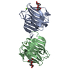
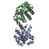
| Remark 300 | BIOMOLECULE: 1 THIS ENTRY CONTAINS THE CRYSTALLOGRAPHIC ASYMMETRIC UNIT WHICH CONSISTS OF 2 CHAIN(S) ...BIOMOLECULE: 1 THIS ENTRY CONTAINS THE CRYSTALLOGRAPHIC ASYMMETRIC UNIT WHICH CONSISTS OF 2 CHAIN(S). These two chains represent the biological dimer, or two biological monomers. |
- Structure visualization
| Structure viewer | Molecule: MolmilJmol/JSmol |
|---|
- Downloads & links
-Download
| PDBx/mmCIF format | 1m08.cif.gz | 70.5 KB | Display | PDBx/mmCIF format |
|---|---|---|---|---|
| PDB format | pdb1m08.ent.gz | 51.5 KB | Display | PDB format |
| PDBx/mmJSON format | 1m08.json.gz | Tree view | PDBx/mmJSON format | |
| Others |  Other downloads Other downloads |
-Validation report
| Arichive directory | https://data.pdbj.org/pub/pdb/validation_reports/m0/1m08ftp://data.pdbj.org/pub/pdb/validation_reports/m0/1m08 | HTTPS FTP |
|---|
-Related structure data
| Related structure data | 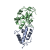 7ceiS S: Starting model for refinement |
|---|---|
| Similar structure data |


-Links
 PDBj
PDBj
- Assembly
| Deposited unit | 
| ||||||||
|---|---|---|---|---|---|---|---|---|---|
| 1 |
| ||||||||
| Unit cell |
|
-Components
| #1: Protein | Mass: 15064.120 Da / Num. of mol.: 2 / Fragment: Nuclease Domain Source method: isolated from a genetically manipulated source Source: (gene. exp.) Escherichia coli str. K12 substr. (bacteria)Species: Escherichia coli / Strain: W3110 / Gene: COLE7 or CEA / Plasmid: pQE70 / Production host: Escherichia coli (E. coli) / Strain (production host): M15References: UniProt: Q47112, Hydrolases; Acting on ester bonds#2: Chemical |   Mass: 65.409 Da / Num. of mol.: 2 / Source method: obtained synthetically / Formula: Zn Mass: 65.409 Da / Num. of mol.: 2 / Source method: obtained synthetically / Formula: Zn#3: Chemical | Phosphate  Mass: 94.971 Da / Num. of mol.: 2 / Source method: obtained synthetically / Formula: PO4 Mass: 94.971 Da / Num. of mol.: 2 / Source method: obtained synthetically / Formula: PO4#4: Water | ChemComp-HOH / | Water Mass: 18.015 Da / Num. of mol.: 224 / Source method: isolated from a natural source / Formula: H2O Mass: 18.015 Da / Num. of mol.: 224 / Source method: isolated from a natural source / Formula: H2O |
|---|
-Experimental details
-Experiment
| Experiment | Method: X-RAY DIFFRACTION / Number of used crystals: 1 |
|---|
- Sample preparation
| Crystal | Density Matthews: 2.4 Å3/Da / Density % sol: 48.5 % | |||||||||||||||||||||||||||||||||||||||||||||||||||||||||||||||
|---|---|---|---|---|---|---|---|---|---|---|---|---|---|---|---|---|---|---|---|---|---|---|---|---|---|---|---|---|---|---|---|---|---|---|---|---|---|---|---|---|---|---|---|---|---|---|---|---|---|---|---|---|---|---|---|---|---|---|---|---|---|---|---|---|
| Crystal grow | Temperature: 298 K / Method: vapor diffusion, hanging drop / pH: 7 Details: sodium phosphate, Sodium Chloride, zinc chloride, ammonium acetate, pH 7.0, VAPOR DIFFUSION, HANGING DROP, temperature 298.0K | |||||||||||||||||||||||||||||||||||||||||||||||||||||||||||||||
| Crystal grow | *PLUS pH: 7.5 | |||||||||||||||||||||||||||||||||||||||||||||||||||||||||||||||
| Components of the solutions | *PLUS
|
-Data collection
| Diffraction | Mean temperature: 120 K |
|---|---|
| Diffraction source | Source: ROTATING ANODE / Type: RIGAKU RU300 / Wavelength: 1.5418 Å |
| Detector | Type: RIGAKU RAXIS II / Detector: IMAGE PLATE / Date: Aug 8, 2001 / Details: mirrors |
| Radiation | Protocol: SINGLE WAVELENGTH / Monochromatic (M) / Laue (L): M / Scattering type: x-ray |
| Radiation wavelength | Wavelength: 1.5418 Å / Relative weight: 1 |
| Reflection | Resolution: 2.1→40 Å / Num. all: 16335 / Num. obs: 16335 / % possible obs: 95 % / Observed criterion σ(F): 0 / Observed criterion σ(I): 0 / Redundancy: 3.3 % / Biso Wilson estimate: 18.3 Å2 / Rsym value: 0.044 / Net I/σ(I): 22.5 |
| Reflection shell | Resolution: 2.1→2.18 Å / Mean I/σ(I) obs: 7 / Num. unique all: 1591 / Rsym value: 0.17 / % possible all: 92.7 |
| Reflection | *PLUS Lowest resolution: 40 Å / % possible obs: 95 % / Num. measured all: 53594 / Rmerge(I) obs: 0.044 |
| Reflection shell | *PLUS % possible obs: 92.7 % / Rmerge(I) obs: 0.173 |
- Processing
| Software |
| ||||||||||||||||||||||||||||||||||||||||||||||||||||||||||||||||||||||||||||||||
|---|---|---|---|---|---|---|---|---|---|---|---|---|---|---|---|---|---|---|---|---|---|---|---|---|---|---|---|---|---|---|---|---|---|---|---|---|---|---|---|---|---|---|---|---|---|---|---|---|---|---|---|---|---|---|---|---|---|---|---|---|---|---|---|---|---|---|---|---|---|---|---|---|---|---|---|---|---|---|---|---|---|
| Refinement | Method to determine structure: MOLECULAR REPLACEMENT Starting model: 7CEI Resolution: 2.1→25.61 Å / Rfactor Rfree error: 0.006 / Data cutoff high absF: 369065.68 / Data cutoff low absF: 0 / Isotropic thermal model: RESTRAINED / Cross valid method: THROUGHOUT / σ(F): 0 / Stereochemistry target values: Engh & Huber
| ||||||||||||||||||||||||||||||||||||||||||||||||||||||||||||||||||||||||||||||||
| Solvent computation | Solvent model: FLAT MODEL / Bsol: 52.3381 Å2 / ksol: 0.370504 e/Å3 | ||||||||||||||||||||||||||||||||||||||||||||||||||||||||||||||||||||||||||||||||
| Displacement parameters | Biso mean: 27.9 Å2
| ||||||||||||||||||||||||||||||||||||||||||||||||||||||||||||||||||||||||||||||||
| Refine analyze |
| ||||||||||||||||||||||||||||||||||||||||||||||||||||||||||||||||||||||||||||||||
| Refinement step | Cycle: LAST / Resolution: 2.1→25.61 Å
| ||||||||||||||||||||||||||||||||||||||||||||||||||||||||||||||||||||||||||||||||
| Refine LS restraints |
| ||||||||||||||||||||||||||||||||||||||||||||||||||||||||||||||||||||||||||||||||
| LS refinement shell | Resolution: 2.1→2.23 Å / Rfactor Rfree error: 0.016 / Total num. of bins used: 6
| ||||||||||||||||||||||||||||||||||||||||||||||||||||||||||||||||||||||||||||||||
| Xplor file |
| ||||||||||||||||||||||||||||||||||||||||||||||||||||||||||||||||||||||||||||||||
| Refinement | *PLUS Highest resolution: 2.1 Å / Lowest resolution: 50 Å / % reflection Rfree: 10 % / Rfactor Rfree: 0.243 / Rfactor Rwork: 0.185 | ||||||||||||||||||||||||||||||||||||||||||||||||||||||||||||||||||||||||||||||||
| Solvent computation | *PLUS | ||||||||||||||||||||||||||||||||||||||||||||||||||||||||||||||||||||||||||||||||
| Displacement parameters | *PLUS | ||||||||||||||||||||||||||||||||||||||||||||||||||||||||||||||||||||||||||||||||
| Refine LS restraints | *PLUS
| ||||||||||||||||||||||||||||||||||||||||||||||||||||||||||||||||||||||||||||||||
| LS refinement shell | *PLUS Lowest resolution: 2.18 Å |
