 Movie
Movie Controller

[English] 日本語
 Yorodumi
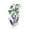
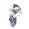
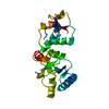
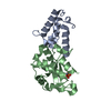
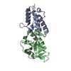
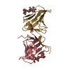
- PDB-1mz8: CRYSTAL STRUCTURES OF THE NUCLEASE DOMAIN OF COLE7/IM7 IN COMPLEX... -
+ Open data

- Basic information
| Entry | Database: PDB / ID: 1mz8 | ||||||
|---|---|---|---|---|---|---|---|
| Title | CRYSTAL STRUCTURES OF THE NUCLEASE DOMAIN OF COLE7/IM7 IN COMPLEX WITH A PHOSPHATE ION AND A ZINC ION | ||||||
 Components Components |
| ||||||
 Keywords Keywords |  Toxin / Hydrolase/protein binding / H-N-H motif / Hydrolase-protein binding COMPLEX Toxin / Hydrolase/protein binding / H-N-H motif / Hydrolase-protein binding COMPLEX | ||||||
| Function / homology |  Function and homology informationextrachromosomal circular DNA / bacteriocin immunity / toxic substance binding / endonuclease activity / killing of cells of another organism / Hydrolases; Acting on ester bonds / defense response to bacterium / metal ion binding Function and homology informationextrachromosomal circular DNA / bacteriocin immunity / toxic substance binding / endonuclease activity / killing of cells of another organism / Hydrolases; Acting on ester bonds / defense response to bacterium / metal ion bindingSimilarity search - Function | ||||||
| Biological species |  Escherichia coli (E. coli) Escherichia coli (E. coli) | ||||||
| Method | X-RAY DIFFRACTION / SYNCHROTRON / MOLECULAR REPLACEMENT / Resolution: 2 Å | ||||||
 Authors Authors | Sui, M.J. / Tsai, L.C. / Hsia, K.C. / Doudeva, L.G. / Ku, W.Y. / Han, G.W. / Yuan, H.S. | ||||||
 Citation Citation | Journal: PROTEIN SCI. / Year: 2002 Title: Metal ions and phosphate binding in the H-N-H motif: crystal structures of the nuclease domain of ColE7/Im7 in complex with a phosphate ion and different divalent metal ions Authors: Sui, M.J. / Tsai, L.C. / Hsia, K.C. / Doudeva, L.G. / Ku, W.Y. / Han, G.W. / Yuan, H.S. | ||||||
| History |
| ||||||
| Remark 300 | BIOMOLECULE: 1, 2 THIS ENTRY CONTAINS THE CRYSTALLOGRAPHIC ASYMMETRIC UNIT WHICH CONSISTS OF 4 ...BIOMOLECULE: 1, 2 THIS ENTRY CONTAINS THE CRYSTALLOGRAPHIC ASYMMETRIC UNIT WHICH CONSISTS OF 4 CHAIN(S), THAT IS TWO HETERODIMERS. SEE REMARK 350 FOR INFORMATION ON GENERATING THE BIOLOGICAL MOLECULE(S). |
- Structure visualization
| Structure viewer | Molecule: MolmilJmol/JSmol |
|---|
- Downloads & links
-Download
| PDBx/mmCIF format | 1mz8.cif.gz | 113 KB | Display | PDBx/mmCIF format |
|---|---|---|---|---|
| PDB format | pdb1mz8.ent.gz | 86.3 KB | Display | PDB format |
| PDBx/mmJSON format | 1mz8.json.gz | Tree view | PDBx/mmJSON format | |
| Others |  Other downloads Other downloads |
-Validation report
| Arichive directory | https://data.pdbj.org/pub/pdb/validation_reports/mz/1mz8ftp://data.pdbj.org/pub/pdb/validation_reports/mz/1mz8 | HTTPS FTP |
|---|
-Related structure data
| Related structure data |  7ceiS S: Starting model for refinement |
|---|---|
| Similar structure data |


-Links
 PDBj
PDBj
- Assembly
| Deposited unit | 
| ||||||||||||
|---|---|---|---|---|---|---|---|---|---|---|---|---|---|
| 1 | 
| ||||||||||||
| 2 | 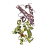
| ||||||||||||
| Unit cell |
| ||||||||||||
| Components on special symmetry positions |
|
-Components
| #1: Protein | Mass: 9906.963 Da / Num. of mol.: 2 Source method: isolated from a genetically manipulated source Source: (gene. exp.) Escherichia coli (E. coli) / Gene: cei7 / Plasmid: pQE70 / Production host: Escherichia coli (E. coli) / Strain (production host): M15 / References: UniProt: Q03708#2: Protein | Mass: 15062.104 Da / Num. of mol.: 2 / Fragment: nuclease domain Source method: isolated from a genetically manipulated source Source: (gene. exp.) Escherichia coli (E. coli) / Gene: cea7 / Plasmid: pQE30 / Production host: Escherichia coli (E. coli) / Strain (production host): M15References: UniProt: Q47112, Hydrolases; Acting on ester bonds#3: Chemical |   Mass: 65.409 Da / Num. of mol.: 2 / Source method: obtained synthetically / Formula: Zn Mass: 65.409 Da / Num. of mol.: 2 / Source method: obtained synthetically / Formula: Zn#4: Chemical | Phosphate  Mass: 94.971 Da / Num. of mol.: 2 / Source method: obtained synthetically / Formula: PO4 Mass: 94.971 Da / Num. of mol.: 2 / Source method: obtained synthetically / Formula: PO4#5: Water | ChemComp-HOH / | Water Mass: 18.015 Da / Num. of mol.: 562 / Source method: isolated from a natural source / Formula: H2O Mass: 18.015 Da / Num. of mol.: 562 / Source method: isolated from a natural source / Formula: H2O |
|---|
-Experimental details
-Experiment
| Experiment | Method: X-RAY DIFFRACTION / Number of used crystals: 1 |
|---|
- Sample preparation
| Crystal | Density Matthews: 2.7 Å3/Da / Density % sol: 54.6 % | ||||||||||||||||||||||||||||||||||||
|---|---|---|---|---|---|---|---|---|---|---|---|---|---|---|---|---|---|---|---|---|---|---|---|---|---|---|---|---|---|---|---|---|---|---|---|---|---|
| Crystal grow | Temperature: 298 K / Method: vapor diffusion, hanging drop / pH: 6.3 Details: PEG4000, sodium phosphate, ammonium acetate, pH 6.3, VAPOR DIFFUSION, HANGING DROP, temperature 298K | ||||||||||||||||||||||||||||||||||||
| Crystal grow | *PLUS | ||||||||||||||||||||||||||||||||||||
| Components of the solutions | *PLUS
|
-Data collection
| Diffraction | Mean temperature: 100 K |
|---|---|
| Diffraction source | Source: SYNCHROTRON / Site: NSRRC  / Beamline: BL17B2 / Wavelength: 1.29004 Å / Beamline: BL17B2 / Wavelength: 1.29004 Å |
| Detector | Type: RIGAKU RAXIS IV / Detector: IMAGE PLATE |
| Radiation | Protocol: SINGLE WAVELENGTH / Monochromatic (M) / Laue (L): M / Scattering type: x-ray |
| Radiation wavelength | Wavelength: 1.29004 Å / Relative weight: 1 |
| Reflection | Resolution: 2→40 Å / Num. all: 169490 / Num. obs: 36897 / % possible obs: 96.1 % / Observed criterion σ(I): 0 / Redundancy: 4.6 % / Biso Wilson estimate: 15.1 Å2 / Rsym value: 0.061 / Net I/σ(I): 21.7 |
| Reflection shell | Resolution: 2→2.07 Å / Mean I/σ(I) obs: 5.5 / Num. unique all: 3430 / Rsym value: 0.278 / % possible all: 96.1 |
| Reflection | *PLUS Num. measured all: 169490 / Rmerge(I) obs: 0.061 |
| Reflection shell | *PLUS Highest resolution: 2 Å / Rmerge(I) obs: 0.278 |
- Processing
| Software |
| ||||||||||||||||||||||||||||||||||||||||||||||||||||||||||||||||||||||||||||||||
|---|---|---|---|---|---|---|---|---|---|---|---|---|---|---|---|---|---|---|---|---|---|---|---|---|---|---|---|---|---|---|---|---|---|---|---|---|---|---|---|---|---|---|---|---|---|---|---|---|---|---|---|---|---|---|---|---|---|---|---|---|---|---|---|---|---|---|---|---|---|---|---|---|---|---|---|---|---|---|---|---|---|
| Refinement | Method to determine structure: MOLECULAR REPLACEMENT Starting model: 7cei Resolution: 2→27.97 Å / Rfactor Rfree error: 0.004 / Data cutoff high absF: 635436.5 / Data cutoff low absF: 0 / Isotropic thermal model: RESTRAINED / Cross valid method: THROUGHOUT / σ(F): 0
| ||||||||||||||||||||||||||||||||||||||||||||||||||||||||||||||||||||||||||||||||
| Solvent computation | Solvent model: FLAT MODEL / Bsol: 62.4188 Å2 / ksol: 0.390765 e/Å3 | ||||||||||||||||||||||||||||||||||||||||||||||||||||||||||||||||||||||||||||||||
| Displacement parameters | Biso mean: 29.7 Å2
| ||||||||||||||||||||||||||||||||||||||||||||||||||||||||||||||||||||||||||||||||
| Refine analyze |
| ||||||||||||||||||||||||||||||||||||||||||||||||||||||||||||||||||||||||||||||||
| Refinement step | Cycle: LAST / Resolution: 2→27.97 Å
| ||||||||||||||||||||||||||||||||||||||||||||||||||||||||||||||||||||||||||||||||
| Refine LS restraints |
| ||||||||||||||||||||||||||||||||||||||||||||||||||||||||||||||||||||||||||||||||
| LS refinement shell | Resolution: 2→2.13 Å / Rfactor Rfree error: 0.011 / Total num. of bins used: 6
| ||||||||||||||||||||||||||||||||||||||||||||||||||||||||||||||||||||||||||||||||
| Xplor file |
| ||||||||||||||||||||||||||||||||||||||||||||||||||||||||||||||||||||||||||||||||
| Refinement | *PLUS Highest resolution: 2 Å / Lowest resolution: 40 Å / % reflection Rfree: 10 % / Rfactor Rfree: 0.241 / Rfactor Rwork: 0.191 | ||||||||||||||||||||||||||||||||||||||||||||||||||||||||||||||||||||||||||||||||
| Solvent computation | *PLUS | ||||||||||||||||||||||||||||||||||||||||||||||||||||||||||||||||||||||||||||||||
| Displacement parameters | *PLUS | ||||||||||||||||||||||||||||||||||||||||||||||||||||||||||||||||||||||||||||||||
| Refine LS restraints | *PLUS
|
