 Movie
Movie Controller

[English] 日本語
 Yorodumi
- PDB-1zea: Structure of the anti-cholera toxin antibody Fab fragment TE33 in... -
+ Open data

- Basic information
| Entry | Database: PDB / ID: 1zea | ||||||
|---|---|---|---|---|---|---|---|
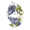
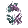
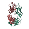
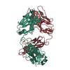
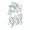
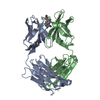
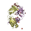
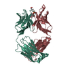
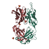
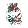
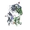
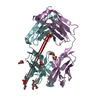
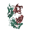
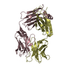
| Title | Structure of the anti-cholera toxin antibody Fab fragment TE33 in complex with a D-peptide | ||||||
 Components Components |
| ||||||
 Keywords Keywords |  IMMUNE SYSTEM / polyspecificity / cross-reactivity / anti-cholera toxin / antigen-antibody complex / antigen recognition / substitution matrix IMMUNE SYSTEM / polyspecificity / cross-reactivity / anti-cholera toxin / antigen-antibody complex / antigen recognition / substitution matrix | ||||||
| Function / homology |  Function and homology information Function and homology information | ||||||
| Biological species |  Mus musculus (house mouse) Mus musculus (house mouse)synthetic construct (others) | ||||||
| Method | X-RAY DIFFRACTION / SYNCHROTRON / MOLECULAR REPLACEMENT / Resolution: 1.78 Å | ||||||
 Authors Authors | Scheerer, P. / Krauss, N. / Wessner, H. / Scholz, C. / Otte, L. / Seifert, M. / Kramer, A. / Schneider-Mergener, J. / Hoehne, W. | ||||||
 Citation Citation | Journal: J.Mol.Recognit. / Year: 2007 Title: Structure of an anti-cholera toxin antibody Fab in complex with an epitope-derived D-peptide: a case of polyspecific recognition. Authors: Scheerer, P. / Kramer, A. / Otte, L. / Seifert, M. / Wessner, H. / Scholz, C. / Krauss, N. / Schneider-Mergener, J. / Hohne, W. | ||||||
| History |
|
- Structure visualization
| Structure viewer | Molecule: MolmilJmol/JSmol |
|---|
- Downloads & links
-Download
| PDBx/mmCIF format | 1zea.cif.gz | 107.6 KB | Display | PDBx/mmCIF format |
|---|---|---|---|---|
| PDB format | pdb1zea.ent.gz | 79.7 KB | Display | PDB format |
| PDBx/mmJSON format | 1zea.json.gz | Tree view | PDBx/mmJSON format | |
| Others |  Other downloads Other downloads |
-Validation report
| Arichive directory | https://data.pdbj.org/pub/pdb/validation_reports/ze/1zeaftp://data.pdbj.org/pub/pdb/validation_reports/ze/1zea | HTTPS FTP |
|---|
-Related structure data
| Related structure data | 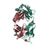 1tetS S: Starting model for refinement |
|---|---|
| Similar structure data |


-Links
 PDBj
PDBj


- Assembly
| Deposited unit | 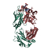
| ||||||||
|---|---|---|---|---|---|---|---|---|---|
| 1 |
| ||||||||
| Unit cell |
|
-Components
| #1: Antibody | Mass: 23843.475 Da / Num. of mol.: 1 / Source method: isolated from a natural source / Source: (natural) Mus musculus (house mouse) / Cell line: Hybridoma / Strain: BALB/c / References: UniProt: A2NHM3 | ||
|---|---|---|---|
| #2: Antibody | Mass: 23410.299 Da / Num. of mol.: 1 / Source method: isolated from a natural source / Source: (natural) Mus musculus (house mouse) / Cell line: Hybridoma / Strain: BALB/c / References: UniProt: Q921A6 | ||
| #3: Protein/peptide | Mass: 990.005 Da / Num. of mol.: 1 / Source method: obtained synthetically Details: This D-amino acid sequence occurs peptide synthesis Source: (synth.) synthetic construct (others) | ||
| #4: Chemical | Citric acid  Mass: 192.124 Da / Num. of mol.: 2 / Source method: obtained synthetically / Formula: C6H8O7 Mass: 192.124 Da / Num. of mol.: 2 / Source method: obtained synthetically / Formula: C6H8O7#5: Water | ChemComp-HOH / | Water Mass: 18.015 Da / Num. of mol.: 276 / Source method: isolated from a natural source / Formula: H2O Mass: 18.015 Da / Num. of mol.: 276 / Source method: isolated from a natural source / Formula: H2O |
-Experimental details
-Experiment
| Experiment | Method: X-RAY DIFFRACTION / Number of used crystals: 1 |
|---|
- Sample preparation
| Crystal | Density Matthews: 2.33 Å3/Da / Density % sol: 46.74 % |
|---|---|
| Crystal grow | Temperature: 293 K / Method: vapor diffusion, hanging drop / pH: 4.5 Details: PEG 8000, citrat buffer, potassium chloride, pH 4.5, VAPOR DIFFUSION, HANGING DROP, temperature 293K |
-Data collection
| Diffraction | Mean temperature: 100 K |
|---|---|
| Diffraction source | Source: SYNCHROTRON / Site: EMBL/DESY, HAMBURG  / Beamline: X13 / Wavelength: 0.8095 Å / Beamline: X13 / Wavelength: 0.8095 Å |
| Detector | Type: MARRESEARCH / Detector: CCD / Date: Oct 26, 2001 / Details: bent mirrors |
| Radiation | Monochromator: Triangular monochromator / Protocol: SINGLE WAVELENGTH / Monochromatic (M) / Laue (L): M / Scattering type: x-ray |
| Radiation wavelength | Wavelength: 0.8095 Å / Relative weight: 1 |
| Reflection | Resolution: 1.78→23 Å / Num. all: 40461 / Num. obs: 40461 / % possible obs: 93.4 % / Observed criterion σ(F): 0 / Observed criterion σ(I): 0 / Redundancy: 4.78 % / Biso Wilson estimate: 20.25 Å2 / Rsym value: 0.064 / Net I/σ(I): 14.14 |
| Reflection shell | Resolution: 1.78→1.81 Å / Redundancy: 3.29 % / Mean I/σ(I) obs: 3.27 / Num. unique all: 1916 / Rsym value: 0.317 / % possible all: 89.4 |
- Processing
| Software |
| |||||||||||||||||||||||||
|---|---|---|---|---|---|---|---|---|---|---|---|---|---|---|---|---|---|---|---|---|---|---|---|---|---|---|
| Refinement | Method to determine structure: MOLECULAR REPLACEMENT Starting model: PDB ENTRY 1TET Resolution: 1.78→23 Å / Isotropic thermal model: isotropic / Cross valid method: THROUGHOUT / σ(F): 0 / σ(I): 0 / Stereochemistry target values: Engh & Huber Details: The current model includes the FAB fragment TE33, residues DVA A 1 - DSN A 9 of the bound D-amino acid peptide D2, 276 water molecules and 2 CITRATE ion. The light and heavy chains have been ...Details: The current model includes the FAB fragment TE33, residues DVA A 1 - DSN A 9 of the bound D-amino acid peptide D2, 276 water molecules and 2 CITRATE ion. The light and heavy chains have been designated *L* and *H*, respectively. Peptide has been designated *A*. The numbering of TE33 residues is essentially according to E. KABAT (E.A. KABAT,T.T. WU, H.M. PERRY, K.S. GOTTESMAN, sequences of proteins of Imuunological interest, 5TH ED.,(1991), NATIONAL INSTITUTES OF HEALTH, BETHESDA, MD.). There is no electron density corresponding to the 7-residue stretch H 127 - H 133 in the constant domain of heavy chain H.
| |||||||||||||||||||||||||
| Displacement parameters | Biso mean: 23.0271 Å2
| |||||||||||||||||||||||||
| Refine analyze |
| |||||||||||||||||||||||||
| Refinement step | Cycle: LAST / Resolution: 1.78→23 Å
| |||||||||||||||||||||||||
| Refine LS restraints |
| |||||||||||||||||||||||||
| LS refinement shell | Resolution: 1.78→1.84 Å / Rfactor Rfree error: 0
|
