 Movie
Movie Controller


+ Open data

- Basic information
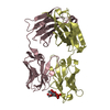
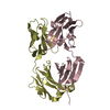
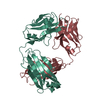
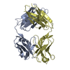
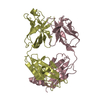
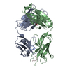
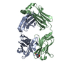
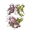
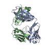
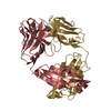
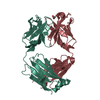
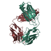
| Entry | Database: PDB / ID: 5ysl | ||||||
|---|---|---|---|---|---|---|---|
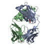
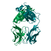
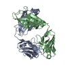
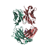
| Title | Crystal structure of antibody 1H1 Fab | ||||||
 Components Components |
| ||||||
 Keywords Keywords |  IMMUNE SYSTEM / Fragment / FAB FRAGMENT / FICIN DIGESTION / INTACT ANTIBODY IgG1 IMMUNE SYSTEM / Fragment / FAB FRAGMENT / FICIN DIGESTION / INTACT ANTIBODY IgG1 | ||||||
| Function / homology | Immunoglobulins / Immunoglobulin-like / Sandwich / Mainly Beta Function and homology information Function and homology information | ||||||
| Biological species |  Mus musculus (house mouse) Mus musculus (house mouse) | ||||||
| Method | X-RAY DIFFRACTION / SYNCHROTRON / MOLECULAR REPLACEMENT / Resolution: 2.5 Å | ||||||
 Authors Authors | Hu, X.L. / Yang, F.L. | ||||||
 Citation Citation | Journal: PLoS Pathog / Year: 2017 Title: Two classes of protective antibodies against Pseudorabies virus variant glycoprotein B: Implications for vaccine design. Authors: Xiangdong Li / Fanli Yang / Xule Hu / Feifei Tan / Jianxun Qi / Ruchao Peng / Min Wang / Yan Chai / Liying Hao / Junhua Deng / Chenyu Bai / Juan Wang / Hao Song / Shuguang Tan / Guangwen Lu ...Authors: Xiangdong Li / Fanli Yang / Xule Hu / Feifei Tan / Jianxun Qi / Ruchao Peng / Min Wang / Yan Chai / Liying Hao / Junhua Deng / Chenyu Bai / Juan Wang / Hao Song / Shuguang Tan / Guangwen Lu / George F Gao / Yi Shi / Kegong Tian /  Abstract: Pseudorabies virus (PRV) belongs to the Herpesviridae family, and is an important veterinary pathogen. Highly pathogenic PRV variants have caused severe epidemics in China since 2011, causing huge ...Pseudorabies virus (PRV) belongs to the Herpesviridae family, and is an important veterinary pathogen. Highly pathogenic PRV variants have caused severe epidemics in China since 2011, causing huge economic losses. To tackle the epidemics, we identified a panel of mouse monoclonal antibodies (mAbs) against PRV glycoprotein B (gB) that effectively block PRV infection. Among these 15 mAbs, fourteen of them block PRV entry in a complement-dependent manner. The remaining one, 1H1 mAb, however can directly neutralize the virus independent of complement and displays broad-spectrum neutralizing activities. We further determined the crystal structure of PRV gB and mapped the epitopes of these antibodies on the structure. Interestingly, all the complement-dependent neutralizing antibodies bind gB at the crown region (domain IV). In contrast, the epitope of 1H1 mAb is located at the bottom of domain I, which includes the fusion loops, indicating 1H1 mAb might neutralize the virus by interfering with the membrane fusion process. Our studies demonstrate that gB contains multiple B-cell epitopes in its crown and base regions and that antibodies targeting different epitopes block virus infection through different mechanisms. These findings would provide important clues for antiviral drug design and vaccine development. | ||||||
| History |
|
- Structure visualization
| Structure viewer | Molecule: MolmilJmol/JSmol |
|---|
- Downloads & links
-Download
| PDBx/mmCIF format | 5ysl.cif.gz | 323.5 KB | Display | PDBx/mmCIF format |
|---|---|---|---|---|
| PDB format | pdb5ysl.ent.gz | 263 KB | Display | PDB format |
| PDBx/mmJSON format | 5ysl.json.gz | Tree view | PDBx/mmJSON format | |
| Others |  Other downloads Other downloads |
-Validation report
| Arichive directory | https://data.pdbj.org/pub/pdb/validation_reports/ys/5yslftp://data.pdbj.org/pub/pdb/validation_reports/ys/5ysl | HTTPS FTP |
|---|
-Related structure data
| Related structure data |  6841C  5ys2C 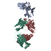 1sy6S S: Starting model for refinement C: citing same article ( |
|---|---|
| Similar structure data |


-Links
 PDBj
PDBj

- Assembly
| Deposited unit | 
| ||||||||
|---|---|---|---|---|---|---|---|---|---|
| 1 | 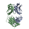
| ||||||||
| 2 | 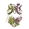
| ||||||||
| 3 | 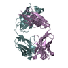
| ||||||||
| 4 | 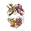
| ||||||||
| Unit cell |
|
-Components
| #1: Antibody | Mass: 23650.258 Da / Num. of mol.: 4 Source method: isolated from a genetically manipulated source Source: (gene. exp.) Mus musculus (house mouse) / Production host:  Homo sapiens (human) Homo sapiens (human)#2: Antibody | Mass: 23666.078 Da / Num. of mol.: 4 Source method: isolated from a genetically manipulated source Source: (gene. exp.) Mus musculus (house mouse) / Production host: Homo sapiens (human) |
|---|
-Experimental details
-Experiment
| Experiment | Method: X-RAY DIFFRACTION / Number of used crystals: 1 |
|---|
- Sample preparation
| Crystal | Density Matthews: 2.41 Å3/Da / Density % sol: 48.97 % |
|---|---|
| Crystal grow | Temperature: 291.15 K / Method: vapor diffusion, sitting drop / pH: 6.8 / Details: 0.2 M potassium sulfate, 20% PEG 3350 |
-Data collection
| Diffraction | Mean temperature: 100 K |
|---|---|
| Diffraction source | Source: SYNCHROTRON / Site: SSRF / Beamline: BL17U1 / Wavelength: 0.97915 Å |
| Detector | Type: ADSC QUANTUM 315 / Detector: CCD / Date: May 12, 2016 |
| Radiation | Protocol: SINGLE WAVELENGTH / Monochromatic (M) / Laue (L): M / Scattering type: x-ray |
| Radiation wavelength | Wavelength: 0.97915 Å / Relative weight: 1 |
| Reflection | Resolution: 2.5→50 Å / Num. obs: 64042 / % possible obs: 98.3 % / Redundancy: 2.9 % / Net I/σ(I): 13.326 |
| Reflection shell | Resolution: 2.5→2.59 Å |
- Processing
| Software |
| ||||||||||||||||||||||||||||||||||||||||||||||||||||||||||||||||||||||||||||||||||||||||||||||||||||||||||||||||||||||||||||||||||||||||||||||||||||||||||||||||||||||||||||||||||||||
|---|---|---|---|---|---|---|---|---|---|---|---|---|---|---|---|---|---|---|---|---|---|---|---|---|---|---|---|---|---|---|---|---|---|---|---|---|---|---|---|---|---|---|---|---|---|---|---|---|---|---|---|---|---|---|---|---|---|---|---|---|---|---|---|---|---|---|---|---|---|---|---|---|---|---|---|---|---|---|---|---|---|---|---|---|---|---|---|---|---|---|---|---|---|---|---|---|---|---|---|---|---|---|---|---|---|---|---|---|---|---|---|---|---|---|---|---|---|---|---|---|---|---|---|---|---|---|---|---|---|---|---|---|---|---|---|---|---|---|---|---|---|---|---|---|---|---|---|---|---|---|---|---|---|---|---|---|---|---|---|---|---|---|---|---|---|---|---|---|---|---|---|---|---|---|---|---|---|---|---|---|---|---|---|
| Refinement | Method to determine structure: MOLECULAR REPLACEMENT Starting model: 1SY6 Resolution: 2.5→50 Å / Cor.coef. Fo:Fc: 0.948 / Cor.coef. Fo:Fc free: 0.921 / SU B: 11.272 / SU ML: 0.248 / Cross valid method: THROUGHOUT / ESU R: 0.646 / ESU R Free: 0.322 / Details: HYDROGENS HAVE BEEN ADDED IN THE RIDING POSITIONS
| ||||||||||||||||||||||||||||||||||||||||||||||||||||||||||||||||||||||||||||||||||||||||||||||||||||||||||||||||||||||||||||||||||||||||||||||||||||||||||||||||||||||||||||||||||||||
| Solvent computation | Ion probe radii: 0.8 Å / Shrinkage radii: 0.8 Å / VDW probe radii: 1.2 Å | ||||||||||||||||||||||||||||||||||||||||||||||||||||||||||||||||||||||||||||||||||||||||||||||||||||||||||||||||||||||||||||||||||||||||||||||||||||||||||||||||||||||||||||||||||||||
| Displacement parameters | Biso mean: 47.266 Å2
| ||||||||||||||||||||||||||||||||||||||||||||||||||||||||||||||||||||||||||||||||||||||||||||||||||||||||||||||||||||||||||||||||||||||||||||||||||||||||||||||||||||||||||||||||||||||
| Refinement step | Cycle: 1 / Resolution: 2.5→50 Å
| ||||||||||||||||||||||||||||||||||||||||||||||||||||||||||||||||||||||||||||||||||||||||||||||||||||||||||||||||||||||||||||||||||||||||||||||||||||||||||||||||||||||||||||||||||||||
| Refine LS restraints |
|