 Movie
Movie Controller

[English] 日本語
 Yorodumi
- PDB-4yny: Crystal structure of monoclonal anti-human podoplanin antibody NZ-1 -
+ Open data

- Basic information
| Entry | Database: PDB / ID: 4yny | |||||||||
|---|---|---|---|---|---|---|---|---|---|---|
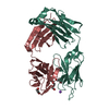
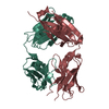
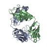
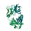
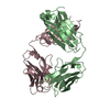
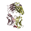
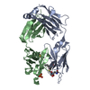
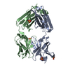
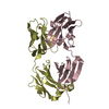
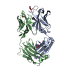
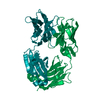
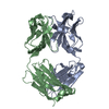
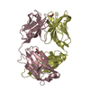
| Title | Crystal structure of monoclonal anti-human podoplanin antibody NZ-1 | |||||||||
 Components Components |
| |||||||||
 Keywords Keywords |  IMMUNE SYSTEM / Antibodies / Monoclonal / Antibody Affinity / Chromatography / Affinity / Epitopes / Rats / Immunoglobulin Fab Fragments / Kinetics / Protein Binding / Proteins / Recombinant Fusion Proteins / Human podoplanin IMMUNE SYSTEM / Antibodies / Monoclonal / Antibody Affinity / Chromatography / Affinity / Epitopes / Rats / Immunoglobulin Fab Fragments / Kinetics / Protein Binding / Proteins / Recombinant Fusion Proteins / Human podoplanin | |||||||||
| Function / homology | Immunoglobulins / Immunoglobulin-like / Sandwich / Mainly Beta Function and homology information Function and homology information | |||||||||
| Biological species |  Rattus norvegicus (Norway rat) Rattus norvegicus (Norway rat) | |||||||||
| Method | X-RAY DIFFRACTION / SYNCHROTRON / MOLECULAR REPLACEMENT / Resolution: 1.584 Å | |||||||||
 Authors Authors | Fujii, Y. / Kitago, Y. / Arimori, T. / Takagi, J. | |||||||||
 Citation Citation | Journal: J.Cell.Sci. / Year: 2016 Title: Tailored placement of a turn-forming PA tag into the structured domain of a protein to probe its conformational state Authors: Fujii, Y. / Matsunaga, Y. / Arimori, T. / Kitago, Y. / Ogasawara, S. / Kaneko, M.K. / Kato, Y. / Takagi, J. | |||||||||
| History |
|
- Structure visualization
| Structure viewer | Molecule: MolmilJmol/JSmol |
|---|
- Downloads & links
-Download
| PDBx/mmCIF format | 4yny.cif.gz | 349.8 KB | Display | PDBx/mmCIF format |
|---|---|---|---|---|
| PDB format | pdb4yny.ent.gz | 284 KB | Display | PDB format |
| PDBx/mmJSON format | 4yny.json.gz | Tree view | PDBx/mmJSON format | |
| Others |  Other downloads Other downloads |
-Validation report
| Arichive directory | https://data.pdbj.org/pub/pdb/validation_reports/yn/4ynyftp://data.pdbj.org/pub/pdb/validation_reports/yn/4yny | HTTPS FTP |
|---|
-Related structure data
| Related structure data |  4yo0C 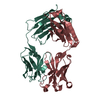 1zanS C: citing same article ( S: Starting model for refinement |
|---|---|
| Similar structure data |


-Links
 PDBj
PDBj

- Assembly
| Deposited unit | 
| ||||||||
|---|---|---|---|---|---|---|---|---|---|
| 1 | 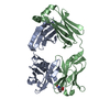
| ||||||||
| 2 | 
| ||||||||
| Unit cell |
|
-Components
| #1: Antibody | Mass: 25560.020 Da / Num. of mol.: 2 Source method: isolated from a genetically manipulated source Source: (gene. exp.) Rattus norvegicus (Norway rat) / Production host: Mus musculus (house mouse)#2: Antibody | Mass: 25494.350 Da / Num. of mol.: 2 Source method: isolated from a genetically manipulated source Source: (gene. exp.) Rattus norvegicus (Norway rat) / Production host: Mus musculus (house mouse)#3: Water | ChemComp-HOH / | Water Mass: 18.015 Da / Num. of mol.: 610 / Source method: isolated from a natural source / Formula: H2O Mass: 18.015 Da / Num. of mol.: 610 / Source method: isolated from a natural source / Formula: H2OSequence details | THE SEQUENCES OF THIS PROTEIN WERE NOT AVAILABLE AT THE UNIPROT KNOWLEDGEBASE DATABASE (UNIPROTKB) ...THE SEQUENCES OF THIS PROTEIN WERE NOT AVAILABLE AT THE UNIPROT KNOWLEDGEB | |
|---|
-Experimental details
-Experiment
| Experiment | Method: X-RAY DIFFRACTION / Number of used crystals: 1 |
|---|
- Sample preparation
| Crystal | Density Matthews: 2.25 Å3/Da / Density % sol: 45.43 % |
|---|---|
| Crystal grow | Temperature: 293 K / Method: vapor diffusion, hanging drop / Details: PEG 4000, sodium citrate |
-Data collection
| Diffraction |
| |||||||||||||||
|---|---|---|---|---|---|---|---|---|---|---|---|---|---|---|---|---|
| Diffraction source |
| |||||||||||||||
| Detector |
| |||||||||||||||
| Radiation |
| |||||||||||||||
| Radiation wavelength |
| |||||||||||||||
| Reflection | Resolution: 1.584→31.5 Å / Num. obs: 118170 / % possible obs: 97.7 % / Redundancy: 6 % / Rmerge(I) obs: 0.13 / Net I/σ(I): 24.5 | |||||||||||||||
| Reflection shell | Highest resolution: 1.584 Å / Redundancy: 3.8 % / Rmerge(I) obs: 0.767 / Mean I/σ(I) obs: 2.3 / % possible all: 89 |


- Processing
| Software |
| |||||||||||||||||||||||||||||||||||||||||||||||||||||||||||||||||||||||||||||||||||||||||||||||||||||||||||||||||||||||||||||||||||||||||||||||||||||||||||||||||||||||||||||||||||||||||||||||||||||||||||||||||||||||||||||||||
|---|---|---|---|---|---|---|---|---|---|---|---|---|---|---|---|---|---|---|---|---|---|---|---|---|---|---|---|---|---|---|---|---|---|---|---|---|---|---|---|---|---|---|---|---|---|---|---|---|---|---|---|---|---|---|---|---|---|---|---|---|---|---|---|---|---|---|---|---|---|---|---|---|---|---|---|---|---|---|---|---|---|---|---|---|---|---|---|---|---|---|---|---|---|---|---|---|---|---|---|---|---|---|---|---|---|---|---|---|---|---|---|---|---|---|---|---|---|---|---|---|---|---|---|---|---|---|---|---|---|---|---|---|---|---|---|---|---|---|---|---|---|---|---|---|---|---|---|---|---|---|---|---|---|---|---|---|---|---|---|---|---|---|---|---|---|---|---|---|---|---|---|---|---|---|---|---|---|---|---|---|---|---|---|---|---|---|---|---|---|---|---|---|---|---|---|---|---|---|---|---|---|---|---|---|---|---|---|---|---|---|---|---|---|---|---|---|---|---|---|---|---|---|---|---|---|---|
| Refinement | Method to determine structure: MOLECULAR REPLACEMENT Starting model: 1ZAN Resolution: 1.584→31.486 Å / SU ML: 0.18 / Cross valid method: THROUGHOUT / σ(F): 1.96 / Phase error: 21.74
| |||||||||||||||||||||||||||||||||||||||||||||||||||||||||||||||||||||||||||||||||||||||||||||||||||||||||||||||||||||||||||||||||||||||||||||||||||||||||||||||||||||||||||||||||||||||||||||||||||||||||||||||||||||||||||||||||
| Solvent computation | Shrinkage radii: 0.9 Å / VDW probe radii: 1.11 Å | |||||||||||||||||||||||||||||||||||||||||||||||||||||||||||||||||||||||||||||||||||||||||||||||||||||||||||||||||||||||||||||||||||||||||||||||||||||||||||||||||||||||||||||||||||||||||||||||||||||||||||||||||||||||||||||||||
| Refinement step | Cycle: LAST / Resolution: 1.584→31.486 Å
| |||||||||||||||||||||||||||||||||||||||||||||||||||||||||||||||||||||||||||||||||||||||||||||||||||||||||||||||||||||||||||||||||||||||||||||||||||||||||||||||||||||||||||||||||||||||||||||||||||||||||||||||||||||||||||||||||
| Refine LS restraints |
| |||||||||||||||||||||||||||||||||||||||||||||||||||||||||||||||||||||||||||||||||||||||||||||||||||||||||||||||||||||||||||||||||||||||||||||||||||||||||||||||||||||||||||||||||||||||||||||||||||||||||||||||||||||||||||||||||
| LS refinement shell |
| |||||||||||||||||||||||||||||||||||||||||||||||||||||||||||||||||||||||||||||||||||||||||||||||||||||||||||||||||||||||||||||||||||||||||||||||||||||||||||||||||||||||||||||||||||||||||||||||||||||||||||||||||||||||||||||||||
| Refinement TLS params. | Method: refined / Refine-ID: X-RAY DIFFRACTION
| |||||||||||||||||||||||||||||||||||||||||||||||||||||||||||||||||||||||||||||||||||||||||||||||||||||||||||||||||||||||||||||||||||||||||||||||||||||||||||||||||||||||||||||||||||||||||||||||||||||||||||||||||||||||||||||||||
| Refinement TLS group |
|
