 Movie
Movie Controller

[English] 日本語
 Yorodumi
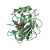
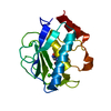
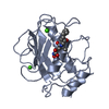
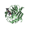
- PDB-1yck: Crystal structure of human peptidoglycan recognition protein (PGRP-S) -
+ Open data

- Basic information
| Entry | Database: PDB / ID: 1yck | ||||||
|---|---|---|---|---|---|---|---|
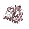
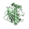
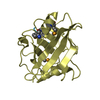
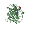
| Title | Crystal structure of human peptidoglycan recognition protein (PGRP-S) | ||||||
 Components Components | Peptidoglycan recognition protein | ||||||
 Keywords Keywords | IMMUNE SYSTEM / PGRP-S / peptidoglycan recognition / innate immunity / pattern recognition proteins | ||||||
| Function / homology |  Function and homology information Function and homology informationpeptidoglycan immune receptor activity / neutrophil degranulation / phagocytic vesicle lumen / N-acetylmuramoyl-L-alanine amidase activity / peptidoglycan binding / antimicrobial humoral response / Antimicrobial peptides / detection of bacterium / peptidoglycan catabolic process / specific granule lumen ...peptidoglycan immune receptor activity / neutrophil degranulation / phagocytic vesicle lumen / N-acetylmuramoyl-L-alanine amidase activity / peptidoglycan binding / antimicrobial humoral response / Antimicrobial peptides / detection of bacterium / peptidoglycan catabolic process / specific granule lumen / antimicrobial humoral immune response mediated by antimicrobial peptide / tertiary granule lumen / killing of cells of another organism / defense response to Gram-positive bacterium / innate immune response / Neutrophil degranulation / extracellular exosome / zinc ion binding / extracellular regionSimilarity search - Function | ||||||
| Biological species |  Homo sapiens (human) Homo sapiens (human) | ||||||
| Method | X-RAY DIFFRACTION / MOLECULAR REPLACEMENT / Resolution: 1.7 Å | ||||||
 Authors Authors | Guan, R. / Wang, Q. / Sundberg, E.J. / Mariuzza, R.A. | ||||||
 Citation Citation | Journal: J.Mol.Biol. / Year: 2005 Title: Crystal structure of human peptidoglycan recognition protein S (PGRP-S) at 1.70 A resolution. Authors: Guan, R. / Wang, Q. / Sundberg, E.J. / Mariuzza, R.A. | ||||||
| History |
|
- Structure visualization
| Structure viewer | Molecule: MolmilJmol/JSmol |
|---|
- Downloads & links
-Download
| PDBx/mmCIF format | 1yck.cif.gz | 50 KB | Display | PDBx/mmCIF format |
|---|---|---|---|---|
| PDB format | pdb1yck.ent.gz | 34.9 KB | Display | PDB format |
| PDBx/mmJSON format | 1yck.json.gz | Tree view | PDBx/mmJSON format | |
| Others |  Other downloads Other downloads |
-Validation report
| Arichive directory | https://data.pdbj.org/pub/pdb/validation_reports/yc/1yckftp://data.pdbj.org/pub/pdb/validation_reports/yc/1yck | HTTPS FTP |
|---|
-Related structure data
| Related structure data |  1sk3S S: Starting model for refinement |
|---|---|
| Similar structure data |


-Links
 PDBj
PDBj

- Assembly
| Deposited unit | 
| |||||||||
|---|---|---|---|---|---|---|---|---|---|---|
| 1 |
| |||||||||
| Unit cell |
| |||||||||
| Components on special symmetry positions |
|
-Components
| #1: Protein | / peptidoglycan recognition protein S / SBBI68 / PGRP-S / UNQ639/PRO1269 Mass: 19456.941 Da / Num. of mol.: 1 Source method: isolated from a genetically manipulated source Source: (gene. exp.) Homo sapiens (human) / Plasmid: pT7-7 / Production host:  Escherichia coli (E. coli) / References: UniProt: O75594 Escherichia coli (E. coli) / References: UniProt: O75594 |
|---|---|
| #2: Water | ChemComp-HOH / Water Mass: 18.015 Da / Num. of mol.: 200 / Source method: isolated from a natural source / Formula: H2O Mass: 18.015 Da / Num. of mol.: 200 / Source method: isolated from a natural source / Formula: H2O |
-Experimental details
-Experiment
| Experiment | Method: X-RAY DIFFRACTION / Number of used crystals: 4 |
|---|
- Sample preparation
| Crystal | Density Matthews: 2.04 Å3/Da / Density % sol: 39.8 % |
|---|---|
| Crystal grow | Temperature: 293 K / Method: vapor diffusion, hanging drop / pH: 5.6 Details: PEG 4000, Sodium Citrate, Sodium Acetate, pH 5.6, VAPOR DIFFUSION, HANGING DROP, temperature 293K |
-Data collection
| Diffraction | Mean temperature: 100 K |
|---|---|
| Diffraction source | Source: ROTATING ANODE / Type: RIGAKU / Wavelength: 1.5418 |
| Detector | Type: RIGAKU RAXIS IV / Detector: IMAGE PLATE / Date: May 6, 2004 |
| Radiation | Monochromator: Si 111 CHANNEL / Protocol: SINGLE WAVELENGTH / Monochromatic (M) / Laue (L): M / Scattering type: x-ray |
| Radiation wavelength | Wavelength: 1.5418 Å / Relative weight: 1 |
| Reflection | Resolution: 1.65→40 Å / Num. all: 19042 / Num. obs: 19042 / % possible obs: 99.8 % / Observed criterion σ(F): 0 / Observed criterion σ(I): 0 / Biso Wilson estimate: 20.3 Å2 / Rmerge(I) obs: 0.067 |
| Reflection shell | Resolution: 1.65→1.71 Å / Redundancy: 6.5 % / Rmerge(I) obs: 0.246 / Mean I/σ(I) obs: 6.4 / Num. unique all: 1090 / % possible all: 100 |
- Processing
| Software |
| |||||||||||||||||||||||||
|---|---|---|---|---|---|---|---|---|---|---|---|---|---|---|---|---|---|---|---|---|---|---|---|---|---|---|
| Refinement | Method to determine structure: MOLECULAR REPLACEMENT Starting model: PDB entry 1SK3 Resolution: 1.7→39.87 Å / Rfactor Rfree error: 0.007 / Data cutoff high absF: 1170490.79 / Data cutoff low absF: 0 / Isotropic thermal model: RESTRAINED / Cross valid method: THROUGHOUT / σ(F): 0 / σ(I): 0 / Stereochemistry target values: Engh & Huber
| |||||||||||||||||||||||||
| Solvent computation | Solvent model: FLAT MODEL / Bsol: 39.8465 Å2 / ksol: 0.361298 e/Å3 | |||||||||||||||||||||||||
| Displacement parameters | Biso mean: 22.3 Å2
| |||||||||||||||||||||||||
| Refine analyze |
| |||||||||||||||||||||||||
| Refinement step | Cycle: LAST / Resolution: 1.7→39.87 Å
| |||||||||||||||||||||||||
| Refine LS restraints |
| |||||||||||||||||||||||||
| LS refinement shell | Resolution: 1.7→1.81 Å / Rfactor Rfree error: 0.022 / Total num. of bins used: 6
| |||||||||||||||||||||||||
| Xplor file |
|
