 Movie
Movie Controller


+ Open data

- Basic information
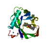
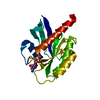
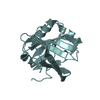
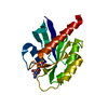
| Entry | Database: PDB / ID: 1sxr | ||||||
|---|---|---|---|---|---|---|---|
| Title | Drosophila Peptidoglycan Recognition Protein (PGRP)-SA | ||||||
 Components Components | Peptidoglycan recognition protein SA CG11709-PA | ||||||
 Keywords Keywords |  IMMUNE SYSTEM / Pattern Recognition Receptor / peptidoglycan / innate immunity / Toll pathway IMMUNE SYSTEM / Pattern Recognition Receptor / peptidoglycan / innate immunity / Toll pathway | ||||||
| Function / homology |  Function and homology information Function and homology informationToll receptor ligand protein activation cascade / Antimicrobial peptides / muramyl dipeptide binding / positive regulation of biosynthetic process of antibacterial peptides active against Gram-positive bacteria / muramoyltetrapeptide carboxypeptidase activity / muramoyltetrapeptide carboxypeptidase / peptidoglycan immune receptor activity / response to peptidoglycan / Neutrophil degranulation / peptidoglycan binding ...Toll receptor ligand protein activation cascade / Antimicrobial peptides / muramyl dipeptide binding / positive regulation of biosynthetic process of antibacterial peptides active against Gram-positive bacteria / muramoyltetrapeptide carboxypeptidase activity / muramoyltetrapeptide carboxypeptidase / peptidoglycan immune receptor activity / response to peptidoglycan / Neutrophil degranulation / peptidoglycan binding / pattern recognition receptor activity / peptidoglycan catabolic process / defense response to Gram-positive bacterium / innate immune response / proteolysis / zinc ion binding / extracellular regionSimilarity search - Function | ||||||
| Biological species |  Drosophila melanogaster (fruit fly) Drosophila melanogaster (fruit fly) | ||||||
| Method | X-RAY DIFFRACTION / SYNCHROTRON / MOLECULAR REPLACEMENT / Resolution: 1.56 Å | ||||||
 Authors Authors | Reiser, J.B. / Teyton, L. / Wilson, I.A. | ||||||
 Citation Citation | Journal: J.Mol.Biol. / Year: 2004 Title: Crystal structure of the Drosophila peptidoglycan recognition protein (PGRP)-SA at 1.56 A resolution Authors: Reiser, J.B. / Teyton, L. / Wilson, I.A. #1: Journal: Mol.Immunol. / Year: 2003Title: CRYSTAL STRUCTURE OF PEPTIDOGLYCAN RECOGNITION PROTEIN LB FROM DROSOPHILA MELANOGASTER Authors: Kim, M.S. / Byun, M. / Oh, B.H. | ||||||
| History |
|
- Structure visualization
| Structure viewer | Molecule: MolmilJmol/JSmol |
|---|
- Downloads & links
-Download
| PDBx/mmCIF format | 1sxr.cif.gz | 90.3 KB | Display | PDBx/mmCIF format |
|---|---|---|---|---|
| PDB format | pdb1sxr.ent.gz | 67.9 KB | Display | PDB format |
| PDBx/mmJSON format | 1sxr.json.gz | Tree view | PDBx/mmJSON format | |
| Others |  Other downloads Other downloads |
-Validation report
| Arichive directory | https://data.pdbj.org/pub/pdb/validation_reports/sx/1sxrftp://data.pdbj.org/pub/pdb/validation_reports/sx/1sxr | HTTPS FTP |
|---|
-Related structure data
| Related structure data |  1ohtS S: Starting model for refinement |
|---|---|
| Similar structure data |


-Links
 PDBj
PDBj

- Assembly
| Deposited unit | 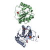
| ||||||||
|---|---|---|---|---|---|---|---|---|---|
| 1 | 
| ||||||||
| 2 | 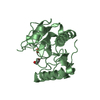
| ||||||||
| 3 | 
| ||||||||
| Unit cell |
| ||||||||
| Components on special symmetry positions |
|
-Components
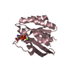
| #1: Protein | Mass: 20295.773 Da / Num. of mol.: 2 Source method: isolated from a genetically manipulated source Source: (gene. exp.) Drosophila melanogaster (fruit fly) / Plasmid: pRMHa3 / Cell (production host): SC2 / Production host: Drosophila melanogaster (fruit fly) / References: UniProt: Q9VYX7#2: Chemical | Sulfate  Mass: 96.063 Da / Num. of mol.: 2 / Source method: obtained synthetically / Formula: SO4 Mass: 96.063 Da / Num. of mol.: 2 / Source method: obtained synthetically / Formula: SO4#3: Chemical | ChemComp-EDO / Ethylene glycol  Mass: 62.068 Da / Num. of mol.: 4 / Source method: obtained synthetically / Formula: C2H6O2 Mass: 62.068 Da / Num. of mol.: 4 / Source method: obtained synthetically / Formula: C2H6O2#4: Water | ChemComp-HOH / | Water Mass: 18.015 Da / Num. of mol.: 388 / Source method: isolated from a natural source / Formula: H2O Mass: 18.015 Da / Num. of mol.: 388 / Source method: isolated from a natural source / Formula: H2O |
|---|
-Experimental details
-Experiment
| Experiment | Method: X-RAY DIFFRACTION / Number of used crystals: 1 |
|---|
- Sample preparation
| Crystal | Density Matthews: 2.55 Å3/Da / Density % sol: 51.43 % |
|---|---|
| Crystal grow | Temperature: 295 K / Method: vapor diffusion, sitting drop / pH: 6 Details: 1.3 to 1.5 M Li2SO4 and 0.1 M MES , pH 6.0, VAPOR DIFFUSION, SITTING DROP, temperature 295K |
-Data collection
| Diffraction | Mean temperature: 100 K |
|---|---|
| Diffraction source | Source: SYNCHROTRON / Site: ALS  / Beamline: 8.2.1 / Wavelength: 1.069 Å / Beamline: 8.2.1 / Wavelength: 1.069 Å |
| Detector | Type: ADSC QUANTUM 210 / Detector: CCD / Date: Sep 13, 2003 |
| Radiation | Protocol: SINGLE WAVELENGTH / Monochromatic (M) / Laue (L): M / Scattering type: x-ray |
| Radiation wavelength | Wavelength: 1.069 Å / Relative weight: 1 |
| Reflection | Resolution: 1.56→45.13 Å / Num. all: 54845 / Num. obs: 54845 / % possible obs: 99.4 % / Observed criterion σ(F): 0 / Observed criterion σ(I): 0 / Redundancy: 3.6 % / Biso Wilson estimate: 17.4 Å2 / Rmerge(I) obs: 0.056 / Rsym value: 0.056 / Net I/σ(I): 10.7 |
| Reflection shell | Resolution: 1.56→1.62 Å / Redundancy: 2.5 % / Rmerge(I) obs: 0.451 / Mean I/σ(I) obs: 2 / Num. unique all: 5148 / Rsym value: 0.451 / % possible all: 94 |
- Processing
| Software |
| |||||||||||||||||||||||||||||||||||||||||||||||||||||||||||||||||||||||||||||||||||||
|---|---|---|---|---|---|---|---|---|---|---|---|---|---|---|---|---|---|---|---|---|---|---|---|---|---|---|---|---|---|---|---|---|---|---|---|---|---|---|---|---|---|---|---|---|---|---|---|---|---|---|---|---|---|---|---|---|---|---|---|---|---|---|---|---|---|---|---|---|---|---|---|---|---|---|---|---|---|---|---|---|---|---|---|---|---|---|
| Refinement | Method to determine structure: MOLECULAR REPLACEMENT Starting model: PDB ENTRY 1OHT Resolution: 1.56→45 Å / Cor.coef. Fo:Fc: 0.962 / Cor.coef. Fo:Fc free: 0.947 / SU B: 1.427 / SU ML: 0.052 / Cross valid method: THROUGHOUT / σ(F): 0 / σ(I): 0 / ESU R: 0.088 / ESU R Free: 0.09 / Stereochemistry target values: Engh & Huber / Details: SSBOND were refined as partially reduced
| |||||||||||||||||||||||||||||||||||||||||||||||||||||||||||||||||||||||||||||||||||||
| Solvent computation | Ion probe radii: 0.8 Å / Shrinkage radii: 0.8 Å / VDW probe radii: 1.2 Å / Solvent model: BABINET MODEL WITH MASK | |||||||||||||||||||||||||||||||||||||||||||||||||||||||||||||||||||||||||||||||||||||
| Displacement parameters | Biso mean: 18.4 Å2
| |||||||||||||||||||||||||||||||||||||||||||||||||||||||||||||||||||||||||||||||||||||
| Refinement step | Cycle: LAST / Resolution: 1.56→45 Å
| |||||||||||||||||||||||||||||||||||||||||||||||||||||||||||||||||||||||||||||||||||||
| Refine LS restraints |
| |||||||||||||||||||||||||||||||||||||||||||||||||||||||||||||||||||||||||||||||||||||
| LS refinement shell | Resolution: 1.56→1.6 Å / Total num. of bins used: 20
|
