 Movie
Movie Controller


+ Open data

- Basic information
| Entry | Database: PDB / ID: 1cxv | ||||||
|---|---|---|---|---|---|---|---|
| Title | STRUCTURE OF RECOMBINANT MOUSE COLLAGENASE-3 (MMP-13) | ||||||
 Components Components | PROTEIN (COLLAGENASE-3) | ||||||
 Keywords Keywords |  HYDROLASE / METALLOPROTEASE / GLYCOPROTEIN / COLLAGEN DEGRADATION HYDROLASE / METALLOPROTEASE / GLYCOPROTEIN / COLLAGEN DEGRADATION | ||||||
| Function / homology |  Function and homology information Function and homology informationpositive regulation of pancreatic trypsinogen secretion / Activation of Matrix Metalloproteinases / Collagen degradation / Assembly of collagen fibrils and other multimeric structures / protein metabolic process => GO:0019538 / growth plate cartilage development / Degradation of the extracellular matrix / endochondral ossification / intercellular canaliculus / Hydrolases; Acting on peptide bonds (peptidases); Metalloendopeptidases ...positive regulation of pancreatic trypsinogen secretion / Activation of Matrix Metalloproteinases / Collagen degradation / Assembly of collagen fibrils and other multimeric structures / protein metabolic process => GO:0019538 / growth plate cartilage development / Degradation of the extracellular matrix / endochondral ossification / intercellular canaliculus / Hydrolases; Acting on peptide bonds (peptidases); Metalloendopeptidases / bone morphogenesis / cartilage development / low-density lipoprotein particle receptor binding / peptide catabolic process / bone mineralization / fibronectin binding / collagen catabolic process / extracellular matrix disassembly / collagen binding / extracellular matrix organization / extracellular matrix / response to hormone / metalloendopeptidase activity / calcium-dependent protein binding / heart development / peptidase activity / endopeptidase activity / lysosome / calcium ion binding / Golgi apparatus / proteolysis / extracellular space / zinc ion binding / extracellular regionSimilarity search - Function | ||||||
| Biological species |  Mus musculus (house mouse) Mus musculus (house mouse) | ||||||
| Method | X-RAY DIFFRACTION / Resolution: 2 Å | ||||||
 Authors Authors | Botos, I. / Meyer, E. / Swanson, S.M. / Lemaitre, V. / Eeckhout, Y. / Meyer, E.F. | ||||||
 Citation Citation | Journal: J.Mol.Biol. / Year: 1999 Title: Structure of recombinant mouse collagenase-3 (MMP-13). Authors: Botos, I. / Meyer, E. / Swanson, S.M. / Lemaitre, V. / Eeckhout, Y. / Meyer, E.F. #1: Journal: Nat.Struct.Biol. / Year: 1999Title: Crystal Structures of Mmp-1 and-13 Reveal the Structural Basis for Selectivity of Collagenase Inhibitors Authors: Lovejoy, B. / Welch, A.R. / Carr, S. / Luong, C. / Broka, C. / Hendriks, T. / Campbell, J.A. / Walker, K.A.M. / Martin, R. / Van, W.H. / Browner, M.F. #2: Journal: Biochem.Biophys.Res.Commun. / Year: 1997Title: The Recombinant Catalytic Domain of Mouse Collagenase-3 Depolymerizes Type I Collagen by Cleaving its Aminotelopeptides Authors: Lemaitre, V. / Jungbluth, A. / Eeckhout, Y. | ||||||
| History |
|
- Structure visualization
| Structure viewer | Molecule: MolmilJmol/JSmol |
|---|
- Downloads & links
-Download
| PDBx/mmCIF format | 1cxv.cif.gz | 88 KB | Display | PDBx/mmCIF format |
|---|---|---|---|---|
| PDB format | pdb1cxv.ent.gz | 65.8 KB | Display | PDB format |
| PDBx/mmJSON format | 1cxv.json.gz | Tree view | PDBx/mmJSON format | |
| Others |  Other downloads Other downloads |
-Validation report
| Arichive directory | https://data.pdbj.org/pub/pdb/validation_reports/cx/1cxvftp://data.pdbj.org/pub/pdb/validation_reports/cx/1cxv | HTTPS FTP |
|---|
-Related structure data
| Similar structure data |
|---|


-Links
 PDBj
PDBj


- Assembly
| Deposited unit | 
| ||||||||
|---|---|---|---|---|---|---|---|---|---|
| 1 | 
| ||||||||
| 2 | 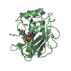
| ||||||||
| Unit cell |
|
-Components
| #1: Protein | Mass: 18520.551 Da / Num. of mol.: 2 / Fragment: CATALYTIC DOMAIN / Source method: isolated from a natural source / Source: (natural) Mus musculus (house mouse)References: UniProt: P33435, Hydrolases; Acting on peptide bonds (peptidases); Metalloendopeptidases#2: Chemical | ChemComp-ZN /   Mass: 65.409 Da / Num. of mol.: 4 / Source method: obtained synthetically / Formula: Zn Mass: 65.409 Da / Num. of mol.: 4 / Source method: obtained synthetically / Formula: Zn#3: Chemical | ChemComp-CA /   Mass: 40.078 Da / Num. of mol.: 4 / Source method: obtained synthetically / Formula: Ca Mass: 40.078 Da / Num. of mol.: 4 / Source method: obtained synthetically / Formula: Ca#4: Chemical |   Mass: 425.883 Da / Num. of mol.: 2 / Source method: obtained synthetically / Formula: C19H20ClNO6S Mass: 425.883 Da / Num. of mol.: 2 / Source method: obtained synthetically / Formula: C19H20ClNO6S#5: Water | ChemComp-HOH / | Water Mass: 18.015 Da / Num. of mol.: 321 / Source method: isolated from a natural source / Formula: H2O Mass: 18.015 Da / Num. of mol.: 321 / Source method: isolated from a natural source / Formula: H2O |
|---|
-Experimental details
-Experiment
| Experiment | Method: X-RAY DIFFRACTION |
|---|
- Sample preparation
| Crystal | Density Matthews: 2.62 Å3/Da / Density % sol: 53.12 % | ||||||||||||||||||||||||
|---|---|---|---|---|---|---|---|---|---|---|---|---|---|---|---|---|---|---|---|---|---|---|---|---|---|
| Crystal grow | *PLUS Temperature: 18 ℃ / pH: 7.5 / Method: batch method | ||||||||||||||||||||||||
| Components of the solutions | *PLUS
|
-Data collection
| Radiation | Protocol: SINGLE WAVELENGTH / Monochromatic (M) / Laue (L): M / Scattering type: x-ray |
|---|---|
| Radiation wavelength | Relative weight: 1 |
| Reflection | Biso Wilson estimate: 14.8 Å2 |
| Reflection | *PLUS Highest resolution: 2 Å / Lowest resolution: 30 Å / Num. obs: 25433 / % possible obs: 96.8 % / Num. measured all: 165387 / Rmerge(I) obs: 0.075 |
| Reflection shell | *PLUS Highest resolution: 2 Å / Lowest resolution: 2.07 Å / % possible obs: 94.3 % / Rmerge(I) obs: 0.286 / Mean I/σ(I) obs: 2.8 |
- Processing
| Software | Name: CNS / Version: 0.5 / Classification: refinement | ||||||||||||||||||||||||||||||||||||||||||||||||||||||||||||||||||||||||||||||||
|---|---|---|---|---|---|---|---|---|---|---|---|---|---|---|---|---|---|---|---|---|---|---|---|---|---|---|---|---|---|---|---|---|---|---|---|---|---|---|---|---|---|---|---|---|---|---|---|---|---|---|---|---|---|---|---|---|---|---|---|---|---|---|---|---|---|---|---|---|---|---|---|---|---|---|---|---|---|---|---|---|---|
| Refinement | Resolution: 2→30 Å / Rfactor Rfree error: 0.005 / Data cutoff high absF: 490456.23 / Data cutoff low absF: 0 / Isotropic thermal model: RESTRAINED / Cross valid method: THROUGHOUT / σ(F): 2
| ||||||||||||||||||||||||||||||||||||||||||||||||||||||||||||||||||||||||||||||||
| Solvent computation | Solvent model: FLAT MODEL / Bsol: 35.88 Å2 / ksol: 0.327 e/Å3 | ||||||||||||||||||||||||||||||||||||||||||||||||||||||||||||||||||||||||||||||||
| Displacement parameters | Biso mean: 28.8 Å2
| ||||||||||||||||||||||||||||||||||||||||||||||||||||||||||||||||||||||||||||||||
| Refine analyze |
| ||||||||||||||||||||||||||||||||||||||||||||||||||||||||||||||||||||||||||||||||
| Refinement step | Cycle: LAST / Resolution: 2→30 Å
| ||||||||||||||||||||||||||||||||||||||||||||||||||||||||||||||||||||||||||||||||
| Refine LS restraints |
| ||||||||||||||||||||||||||||||||||||||||||||||||||||||||||||||||||||||||||||||||
| Refine LS restraints NCS | NCS model details: CONSTR | ||||||||||||||||||||||||||||||||||||||||||||||||||||||||||||||||||||||||||||||||
| LS refinement shell | Resolution: 2→2.13 Å / Rfactor Rfree error: 0.014 / Total num. of bins used: 6
| ||||||||||||||||||||||||||||||||||||||||||||||||||||||||||||||||||||||||||||||||
| Xplor file |
| ||||||||||||||||||||||||||||||||||||||||||||||||||||||||||||||||||||||||||||||||
| Software | *PLUS Version: 0.5 / Classification: refinement | ||||||||||||||||||||||||||||||||||||||||||||||||||||||||||||||||||||||||||||||||
| Refinement | *PLUS σ(F): 2 / % reflection Rfree: 9.9 % | ||||||||||||||||||||||||||||||||||||||||||||||||||||||||||||||||||||||||||||||||
| Solvent computation | *PLUS | ||||||||||||||||||||||||||||||||||||||||||||||||||||||||||||||||||||||||||||||||
| Displacement parameters | *PLUS Biso mean: 28.8 Å2 | ||||||||||||||||||||||||||||||||||||||||||||||||||||||||||||||||||||||||||||||||
| Refine LS restraints | *PLUS
| ||||||||||||||||||||||||||||||||||||||||||||||||||||||||||||||||||||||||||||||||
| LS refinement shell | *PLUS Rfactor Rfree: 0.257 / % reflection Rfree: 10.7 % / Rfactor Rwork: 0.229 |
