 Movie
Movie Controller

[English] 日本語
 Yorodumi
- PDB-1y7q: Mammalian SCAN domain dimer is a domain-swapped homologue of the ... -
+ Open data

- Basic information
| Entry | Database: PDB / ID: 1y7q | ||||||
|---|---|---|---|---|---|---|---|
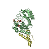
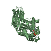
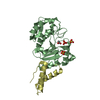
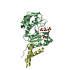
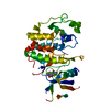
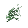
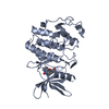
| Title | Mammalian SCAN domain dimer is a domain-swapped homologue of the HIV capsid C-terminal domain | ||||||
 Components Components | Zinc finger protein 174 | ||||||
 Keywords Keywords | TRANSCRIPTION / SCAN domain / retroviral capsid C-terminal domain / dimer / C2H2 zinc finger associated | ||||||
| Function / homology |  Function and homology information Function and homology informationsequence-specific double-stranded DNA binding / actin cytoskeleton / sequence-specific DNA binding / transcription cis-regulatory region binding / DNA-binding transcription factor activity, RNA polymerase II-specific / RNA polymerase II cis-regulatory region sequence-specific DNA binding / DNA-binding transcription factor activity / negative regulation of DNA-templated transcription / chromatin / regulation of transcription by RNA polymerase II ...sequence-specific double-stranded DNA binding / actin cytoskeleton / sequence-specific DNA binding / transcription cis-regulatory region binding / DNA-binding transcription factor activity, RNA polymerase II-specific / RNA polymerase II cis-regulatory region sequence-specific DNA binding / DNA-binding transcription factor activity / negative regulation of DNA-templated transcription / chromatin / regulation of transcription by RNA polymerase II / negative regulation of transcription by RNA polymerase II / protein homodimerization activity / nucleoplasm / metal ion binding / nucleus / plasma membrane / cytosolSimilarity search - Function | ||||||
| Biological species |  Homo sapiens (human) Homo sapiens (human) | ||||||
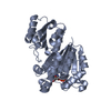
| Method | SOLUTION NMR / distance geometry simulated annealing torsion angle dynamics | ||||||
 Authors Authors | Ivanov, D. / Stone, J.R. / Maki, J.L. / Collins, T. / Wagner, G. | ||||||
 Citation Citation | Journal: Mol.Cell / Year: 2005 Title: Mammalian SCAN Domain Dimer Is a Domain-Swapped Homolog of the HIV Capsid C-Terminal Domain Authors: Ivanov, D. / Stone, J.R. / Maki, J.L. / Collins, T. / Wagner, G. | ||||||
| History |
|
- Structure visualization
| Structure viewer | Molecule: MolmilJmol/JSmol |
|---|
- Downloads & links
-Download
| PDBx/mmCIF format | 1y7q.cif.gz | 1.2 MB | Display | PDBx/mmCIF format |
|---|---|---|---|---|
| PDB format | pdb1y7q.ent.gz | 1 MB | Display | PDB format |
| PDBx/mmJSON format | 1y7q.json.gz | Tree view | PDBx/mmJSON format | |
| Others |  Other downloads Other downloads |
-Validation report
| Arichive directory | https://data.pdbj.org/pub/pdb/validation_reports/y7/1y7qftp://data.pdbj.org/pub/pdb/validation_reports/y7/1y7q | HTTPS FTP |
|---|
-Related structure data
| Similar structure data |
|---|


-Links
 PDBj
PDBj


- Assembly
| Deposited unit | 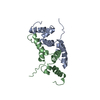
| |||||||||
|---|---|---|---|---|---|---|---|---|---|---|
| 1 |
| |||||||||
| NMR ensembles |
|
-Components
| #1: Protein | / AW-1 / Zinc finger and SCAN domain containing protein 8 Mass: 11626.498 Da / Num. of mol.: 2 / Fragment: SCAN domain, residues 37-132 / Mutation: P100E, P111L Source method: isolated from a genetically manipulated source Source: (gene. exp.) Homo sapiens (human) / Gene: ZNF174, ZSCAN8 / Production host:  Escherichia coli (E. coli) / References: UniProt: Q15697 Escherichia coli (E. coli) / References: UniProt: Q15697 |
|---|
-Experimental details
-Experiment
| Experiment | Method: SOLUTION NMR |
|---|
- Sample preparation
| Details |
| ||||||||||||
|---|---|---|---|---|---|---|---|---|---|---|---|---|---|
| Sample conditions | Ionic strength: 200mM NaCl / pH: 6.5 / Pressure: ambient / Temperature: 298 K |
-NMR measurement
| NMR spectrometer |
|
|---|
- Processing
| NMR software |
| ||||||||||||||||
|---|---|---|---|---|---|---|---|---|---|---|---|---|---|---|---|---|---|
| Refinement | Method: distance geometry simulated annealing torsion angle dynamics Software ordinal: 1 | ||||||||||||||||
| NMR representative | Selection criteria: lowest energy | ||||||||||||||||
| NMR ensemble | Conformer selection criteria: structures with favorable non-bond energy Conformers calculated total number: 100 / Conformers submitted total number: 20 |
